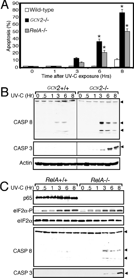
Figure 6. Loss of GCN2 activity enhances apoptosis in response to UV exposure.
GCN2−/− and RelA/p65−/− MEF cells, and their wild-type counterparts, were exposed to 30 J/m2 UV-C irradiation or no stress (0), and incubated in the culture medium for up to 8 h as indicated. (A) Apoptosis was monitored by using the TUNEL assay to detect fragmented DNA. Wild-type GCN2+/+ cells and RelA/p65+/+ gave similar results, and the GCN2+/+ apoptotic measurements are presented. Means values±S.D. were derived from three independent experiments. Two-way ANOVA and post hoc analyses indicated that there were significant differences in apoptosis between genotypes (P<0.001) and with time (P<0.001) as indicated by the asterisks. (B) Cleavage and activation of caspase (CASP) 3 and 8 were monitored by immunoblot analysis using polyclonal antibody specific to each caspase. Arrowheads indicate the non-cleaved caspase 8 (upper band), and cleaved/activated capase 8 (two lower bands). The polyclonal antibody used in the capase 3 immunoblot recognizes only the cleaved, activated caspase 3 polypeptide highlighted by the arrowhead. (C) RelA/p65+/+ and RelA/p65−/− MEF cells were subjected to 30 J/m2 UV-C irradiation or no stress (0), and cultured for between 0.5 and 8 h as indicated. Levels of phosphorylated eIF2α, total eIF2α, actin and cleaved and activated caspase 3 and 8 were measured by immunoblot analysis. Arrowheads indicate the uncleaved and cleaved/activated versions of caspase 8, and activated caspase 3, as described for (B). Immunoblot experiments shown in (B) and (C) are representative of three independent experiments.

