Abstract
Increasingly many studies reveal how ribosome composition can be tuned to optimally translate the transcriptome of individual cell types. In this study, we investigated the expression pattern, structure within the ribosome and effect on protein synthesis of the ribosomal protein paralog 39L (RPL39L). With a novel mass spectrometric approach we revealed the expression of RPL39L protein beyond mouse germ cells, in human pluripotent cells, cancer cell lines and tissue samples. We generated RPL39L knock-out mouse embryonic stem cell (mESC) lines and demonstrated that RPL39L impacts the dynamics of translation, to support the pluripotency and differentiation, spontaneous and along the germ cell lineage. Most differences in protein abundance between WT and RPL39L KO lines were explained by widespread autophagy. By CryoEM analysis of purified RPL39 and RPL39L-containing ribosomes we found that, unlike RPL39, RPL39L has two distinct conformations in the exposed segment of the nascent peptide exit tunnel, creating a distinct hydrophobic patch that has been predicted to support the efficient co-translational folding of alpha helices. Our study shows that ribosomal protein paralogs provide switchable modular components that can tune translation to the protein production needs of individual cell types.
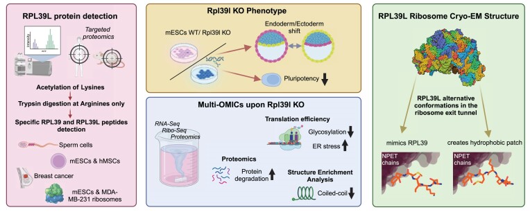
Graphical Abstract
Graphical Abstract.
Introduction
Protein synthesis is carried out by the ribosome, a highly conserved molecular machine with the same basic architecture in all free living organisms. In mammals, the small, 40S ribosomal subunit contains the 18S ribosomal RNA (rRNA) and 33 ribosomal proteins (RPs), while the large 60S subunit contains 46 RPs along with the 5S, 5.8S and 28S rRNAs. Gene duplications gave rise to RP paralogs (1), some with evolutionarily-conserved tissue-specific patterns of expression (2). For instance, Rps27 and Rps27l have both been found to target p53 through their interaction with E3 ubiquitin ligase Mdm2 (3). Rpl22 and Rpl22l have been shown to regulate the splicing of pre-mRNA during morphogenesis, but also to target each other's transcript for degradation, as a simple mechanism to maintain stable protein levels within the cells (4). Rpl3 is necessary for myotube formation and growth (5), while its paralog Rpl3l is important for cardiac muscle contraction (6,7). A particular subset of RP paralogs resulted from the retrotransposition of X-chromosome-located RPs on autosomal chromosomes. These include RPL36AL, RPL10L and RPL39L (8), as well as RPS4 (9). These RPs have strong expression bias for the male germ cell lineage, which would allow them to compensate for their respective X chromosome-encoded paralogs upon meiotic sex chromosome inactivation. Indeed, this function was demonstrated for RPL10L (10). RPL39L is a recently evolved (1) and non-redundant paralog of RPL39L that has just been implicated in the translation of long-lived, sperm cell-specific proteins (11). However, the RPL39L mRNA was also observed outside of the germ cell lineage, particularly in ovarian (12) and breast cancer tissues (2), as well as in lung cancer (13) and neuroblastoma (11) cell lines, where the expression appears to be driven by gene amplifications (14) and CpG island hypomethylation (13). These observations suggest that RPL39L’s function extends beyond the translation of long-lived sperm cell proteins. Unraveling this function has been challenging. RPL39L differs from RPL39 by only 4 or 3 amino acids in human and mouse, respectively, explaining why antibodies that can distinguish RPL39L from RPL39 are still lacking. In addition, the high arginine/lysine content of RPL39L leads to the almost complete digestion of the protein during standard sample preparation for mass spectrometry, probably restricting its detection to cell types with very high expression (11). As pure populations of RPL39L-containing ribosomes have not been obtained so far (11), how RPL39/RPL39L ribosomes differ has also remained unclear.
Our study aimed to determine the role of RPL39/RPL39L ribosome heterogeneity across mammalian cell types. We took advantage of mouse embryonic stem cells (mESC), a cell type with native expression of RPL39L, to generate RPL39L-deficient mESC lines by CRISPR/Cas9 genome editing. We then characterized their gene expression and capacity to differentiate both spontaneously and towards the sperm cell lineage. Ribosome footprinting along with mass spectrometric analysis in the presence and absence of protein degradation inhibitors revealed that RPL39L supports the translation of a heterogeneous collection of proteins. Many of these are involved in cell motility and polarization, thus explaining the critical role of RPL39L in spermatogenesis. As obtaining pure populations of RPL39L ribosomes from mice remains challenging, to analyze the impact of RPL39/RPL39L on ribosome structure we turned to the yeast, which has only the RPL39 gene. We complemented RPL39-deficient yeast cells with either mouse RPL39 or RPL39L, and analyzed purified RPL39 and RPL39L ribosomes. While mouse and yeast RPL39 occupy virtually identical positions in the ribosome exit tunnel, RPL39L was found in two distinct conformations, one very similar and the other distinct from RPL39. The alternative conformation creates a hydrophobic patch in the vestibular region of the nascent peptide exit tunnel (NPET), which was previously postulated to be necessary for the folding of amphipathic ɑ-helices (15). Our results provide an example of paralogous RPs supporting the generation of ribosomes with distinct biophysical properties. In particular, the flexibility conferred to the peptide exit tunnel of the ribosome by RPL39L relative to RPL39 appears to be important for the folding and stability of proteins with long helical domains, many of which are abundant in the male germ cells, but can be more generally described as relevant for cell motility and polarization.
Materials and methods
Quantification of RP gene expression in public RNA-seq data
Raw gene expression counts generated by STAR (16) for all 11 274 samples of the TCGA projects were downloaded from the GDC portal (https://portal.gdc.cancer.gov/). In addition, STAR 2-PASS genomic alignments (BAM format) of short reads from 1226 breast cancer and solid normal tissue samples of the TCGA-BRCA project were obtained from the GDC portal (accession number phs000178.v11.p8). Short reads from a single cells study of embryogenesis (17) were downloaded in .fastq format from the NCBI Sequence Read Archive (SRA, https://www.ncbi.nlm.nih.gov/sra) and aligned with STAR v2.7.10b in 2-PASS mode. Normalized gene TPM values across cell types and tissues estimated from pseudo-bulk scRNA-seq data (https://www.proteinatlas.org/download/rna_single_cell_type_tissue.tsv.zip) and cell-level gene count data (https://www.proteinatlas.org/download/rna_single_cell_read_count.zip) were downloaded from the HPA portal (18).
From 90 RPs expressed in human cells, a subset of 4 core RPs (RPS9, RPS14, RPL4, RPL32) was stringently selected to satisfy the following criteria: (i) RPs are deeply embedded into the rRNA (19), (ii) bind early to ribosomal subunits during synthesis and assembly (20), (iii) are present in other taxonomic groups apart from eukaryotes (21), (iv) do not have strong paralogs in the human genome with DIOPT score >2 and ‘high’ DIOPT rank (22), (v) do not display strong evidence of tissue-specific expression (2). RPs with these features are more likely to be essential for cell survival (Supplementary Figure S1A), as indicated by significantly lower scores in CRISPR (23) and RNAi screening (24) (data available at the DepMap portal https://depmap.org/portal/).
mRNAs encoding RP genes were extracted from the v36 version of the GENCODE comprehensive annotation of the hg38 human genome assembly (25). RNA-SeQC (26) was utilized to obtain raw gene counts from uniquely mapped reads (MAPQ = 255) as well as from all reads, including multi-mapped ones (MAPQ>=0), in bulk RNA-seq data from the TCGA-BRCA project and scRNA-seq data from the embryogenesis study (17). Gene counts of RPL39L and of core RPs comprised only a small fraction (<7%) of multi-mapped reads in breast cancer and normal tissue samples (Supplementary Figure S1B), consistent with these RPs having a relatively low number of processed pseudogenes (27). Gene lengths of the RPs were estimated as average transcript lengths weighted by TPM values. StringTie v.2.2.1 (28) was utilized to identify mRNAs and quantify their TPM values across TCGA-BRCA samples. Median gene lengths of RPL39L, RPL39, and core RPs were checked and found to have only minor differences between cancer and normal samples, indicating minimal isoform variation (Supplementary Figure S1C). The arithmetic means of these median values were further used as gene length estimates.
For RPL39L, RPL39 and core RPs, reads-per-kilobase (RPK) values were estimated from public bulk RNA-seq data from the TCGA project and scRNA-seq data from the embryogenesis study (17) as raw gene counts with pseudocount = 1 divided by the gene length. RPL39-to-core RP and RPL39L-to-core RP ratios were calculated as the ratios of RPL39 (respectively, RPL39L) RPK values relative to the median of core RP RPK values.
Cell culture
In the absence of feeder cells, the WT E14 mESC line was cultured in Dulbecco's Modified Eagle Media (DMEM) (Gibco 31 966), which contained 20% fetal bovine serum (FBS; Gibco 16 141 079) tested for optimal mESC growth, NEAA (Gibco 11 140 050), sodium pyruvate (Gibco 11 360 039), 100 U/mL LIF (Millipore ESG1107) and 0.1 mM 2-ß-mercaptoethanol (Merck ES-007-E), on 0.2% gelatin-coated plates. The culture medium was changed daily, and the cells were passaged every second or third day. Cells were cultured at 37°C in 5% CO2.
RPL39L CRISPR in E14.Tg2a (E14) mouse embryonic stem cells
All transfections for the CRISPR knockout were performed using the lipofectamine 2000 reagent (Life technologies), according to manufacturer's instructions. SgRNAs for the RPL39L gene were designed in pairs from the sequences upstream and downstream of the 5′ and 3′UTRs, respectively. Upstream sgRNAs were cloned into the px330 plasmid backbone with an mCherry marker, and downstream sgRNAs were cloned in the px330 backbone with a GFP marker. The cells positive for both mCherry and GFP expression were FACS-sorted. For single cell colony selection, FACS sorted cells were diluted to a concentration of 0.75 cells per 100 μl in the ESC culture media. 100 μl of this solution was added to each well of a 96-well plate. The cells were allowed to grow for 2 days before wells with single clones were marked. Forward primers for CRISPR KO validation were designed upstream of the upstream sgRNAs and the reverse primers were designed downstream of the downstream sgRNAs (see Supplementary Table S1). In clones with homozygous KO the PCR product should consist of a single band, migrating lower than the wild type band (692 and 587 bp in KO, compared to 1157bp in WT) when run on a 1.3% agarose gel. The bands were excised and gel purified using the Qiagen Gel purification Kit, according to manufacturer's instructions. The purified DNA was cloned into a pUC19 plasmid backbone using Zero Blunt Topo kit (ThermoFischer) according to manufacturer's instructions. This plasmid was sequenced to validate the complete excision of RPL39L.
LC–MS analysis
Sample preparation
Cells were collected and lysed in 50 μl lysis buffer (2 M guanidinium–HCl, 0.1 M HEPES, 5 mM TCEP, pH 8.3) using strong ultra-sonication (10 cycles, Bioruptor, Diagnode). Protein concentration was determined by BCA assay (Thermo Fisher Scientific 23 227) using a small sample aliquot. Sample aliquots containing 50 μg of total proteins were supplemented with lysis buffer to 50 μl, reduced for 10 min at 95°C and alkylated at 10 mM iodoacetamide for 30 min at 25°C followed by adding N-acetyl-cysteine to a final concentration of 12.5 mM to quench any iodoacetamide excess. For global proteomics analyses, proteins were directly digested by incubation with sequencing-grade modified trypsin (1/50, w/w; Promega, Madison, Wisconsin) overnight at 37°C. For targeted analysis of RPL39 and RPL39L, a mixture containing 100 fmol of heavy reference peptides were added to the samples. Then, protein samples were propionylated by adding N-(propionyloxy)-succinimide (0.15 M in DMSO) to a final concentration of 22.5 mM and incubating for 2 h at 25°C with shaking at 500 rpm. This step prevents trypsin from cleaving after lysine residues and leads to the generation of larger peptides suited for LC–MS analysis for our target proteins. To quench the labeling reaction, 1.5 μl aqueous 1.5 M hydroxylamine solution was added and samples were incubated for another 10 min at 25°C shaking at 500 rpm. Subsequently, the pH of the samples was increased to 11.9 by adding 1 M potassium phosphate buffer (pH 12) and incubated for 20 min at 25°C shaking at 500 rpm to remove propionic acid linked to peptide hydroxyl groups. The reaction was stopped by adding 2 M hydrochloric acid until a pH < 2 was reached. After adding 70 μl of a 1 M TEAB (pH 8.5), proteins were digested by incubation with sequencing-grade modified trypsin (1/50, w/w; Promega, Madison, Wisconsin) overnight at 37°C. For all samples, the generated peptides were cleaned up using iST cartridges (PreOmics, Munich, Germany) according to the manufacturer's instructions. Samples were dried under vacuum and stored at −80°C until further use.
Global proteomics LC–MS analysis
Dried peptides were resuspended in 0.1% aqueous formic acid and subjected to LC–MS/MS analysis using a Q Exactive HF Mass Spectrometer fitted with an EASY-nLC 1000 (both Thermo Fisher Scientific) and a custom-made column heater set to 60°C. Peptides were resolved using a RP-HPLC column (75μm × 30cm) packed in-house with C18 resin (ReproSil-Pur C18-AQ, 1.9 μm resin; Dr. Maisch GmbH) at a flow rate of 0.2 μl min–1. The following gradient was used for peptide separation: from 5% B to 15% B over 10 min to 30% B over 60 min to 45% B over 20 min to 95% B over 2 min followed by 18 min at 95% B. Buffer A was 0.1% formic acid in water and buffer B was 80% acetonitrile, 0.1% formic acid in water.
The mass spectrometer was operated in DDA mode with a total cycle time of approximately 1 s. Each MS1 scan was followed by high-collision-dissociation (HCD) of the 10 most abundant precursor ions with dynamic exclusion set to 30 seconds. For MS1, 3e6 ions were accumulated in the Orbitrap over a maximum time of 100 ms and scanned at a resolution of 120 000 FWHM (at 200 m/z). MS2 scans were acquired at a target setting of 1e5 ions, maximum accumulation time of 100 ms and a resolution of 30 000 FWHM (at 200 m/z). Singly charged ions and ions with unassigned charge state were excluded from triggering MS2 events. The normalized collision energy was set to 35%, the mass isolation window was set to 1.1 m/z and one microscan was acquired for each spectrum.
Targeted LC–MS analysis of RPL39L
Parallel reaction-monitoring (PRM) assays (29,30) were generated from a mixture containing 25 fmol/μl of each proteotypic N-(propionyloxy)-succinimid labeled heavy reference peptide (SSHKTFR (RPL39 human and mouse), SSHKTFTIKR (RPL39L human), ASHKTFR (rpl39l mouse), JPT Peptide Technologies GmbH). 2 μl of this standard peptide mix were subjected to LC–MS/MS analysis using a Q Exactive plus Mass Spectrometer fitted with an EASY-nLC 1000 (both Thermo Fisher Scientific) and a custom-made column heater set to 60°C. Peptides were resolved using a EasySpray RP-HPLC column (75 μm × 25 cm, Thermo Fisher Scientific) and a pre-column setup at a flow rate of 0.2 μl/min. The mass spectrometer was operated in DDA mode. Each MS1 scan was followed by high-collision-dissociation (HCD) of the precursor masses of the imported isolation list and the 20 most abundant precursor ions with dynamic exclusion for 20 s. Total cycle time was approximately 1 s. For MS1, 3e6 ions were accumulated in the Orbitrap cell over a maximum time of 50 ms and scanned at a resolution of 70 000 FWHM (at 200 m/z). MS1 triggered MS2 scans were acquired at a target setting of 1e5 ions, a resolution of 17 500 FWHM (at 200 m/z) and a mass isolation window of 1.4 Th. Singly charged ions and ions with unassigned charge state were excluded from triggering MS2 events. The normalized collision energy was set to 27% and one microscan was acquired for each spectrum.
The acquired raw-files were searched using the MaxQuant software (Version 1.6.2.3) against the same human and mouse database mentioned above using default parameters except protein, peptide and site FDR were set to 1 and Lys8, Arg10 and propyl (K) were added as variable modifications. The search results were imported into Skyline (v21.1.0.278) (31) to build a spectral library and assign the most intense transitions to each peptide. An unscheduled mass isolation list containing all peptide ion masses was exported and imported into the Q Exactive Plus operating software for PRM analysis. For PRM-MS analysis, peptide samples were resuspended in 0.1% aqueous formic acid. Due to the required protein propionylation for rpl39 and rpl39l LC–MS analysis, the heavy reference peptides were already spiked in at a concentration of 2 fmol of heavy reference peptides per 1 μg of total endogenous peptide mass during sample preparation (see above). The samples were subjected to LC–MS/MS analysis on the same LC–MS system described above using the following settings: The MS2 resolution of the orbitrap was set to 17 500/140 000 FWHM (at 200 m/z) and the fill time to 50/500 ms for heavy/light peptides. AGC target was set to 3e6, the normalized collision energy was set to 27%, ion isolation window was set to 0.4 m/z and the first mass was fixed to 100 m/z. A MS1 scan at 35 000 resolution (FWHM at 200 m/z), AGC target 3e6 and fill time of 50 ms was included in each MS cycle. All raw-files were imported into Skyline for protein/peptide quantification. To control for sample amount variations during sample preparation, the total ion chromatogram (only comprising precursor ions with two to five charges) of each sample was determined using Progenesis QI software (Nonlinear Dynamics (Waters), Version 2.0) and used for normalization of light (endogenous) peptide abundances.
Sample preparation for inhibition of protein degradation
E14 and RPL39L KO cells were treated for 5 h at 37°C and 5% CO2 with 10 μM (S)-MG132 (STEMCELL Technologies Catalog #73 264) and 5 μM Bafilomycin-A1 (STEMCELL Technologies Catalog #74 242) in the normal media described above. For LC–MS as well as for western blot analysis, cells were scraped and washed twice with warm DPBS, then lysed in a corresponding lysis buffer.
Spontaneous differentiation
Spontaneous differentiation of mESC lines was carried out in conventional mESC medium (as stated above) with 10% fetal bovine serum and no LIF (32). To avoid attachment, embryoid bodies (EBs) were generated by growing 750 000 cells in suspension for 6 days in non-adherent dishes (Greiner Bio-One 633 181). Spheroids were allowed to grow without disturbing the plate for the first 3 days. The medium was changed every second day afterwards. The EBs were harvested after 6 days with 25ml Pasteur pipettes and washed with 1× PBS thrice before further analysis.
Spermatogenic differentiation
Differentiation of mESCs toward sperm cells was done according to the protocol described in ref. (33). In short, E14 cells were harvested and plated as a hanging media drop on Petri dishes filled with PBS on the bottom of the plate, with 1250 ESCs per 25 μl in each drop. The resultant EBs were transferred onto Petri dishes (10–15 EB per dish) after 3 days in hanging drop culture. Neurobasal medium (Gibco 21 103 049), supplemented with B27 (Invitrogen 17 504 044) was used as the differentiation medium, according to the manufacturer's protocol 0.1 μM retinoic acid final concentration (Sigma R2625-50MG) was added to the culture medium one day after EB transfer, and the medium was replaced every two days to minimize deterioration. The contents of each plate were harvested after 4 days.
Polysome profiling and ribo-seq
For ribo-seq analysis, WT and RPL39L KO E14 mESCs were propagated in 5 × 15 cm Petri Dishes (Falcon, 353 025) per sample as described in the cell culture section. The medium was replenished three hours prior to cell collection at the confluency of 50–70%. Before harvesting, the cells were treated with 100μg/ml cycloheximide (CHX) (Sigma, G7698) for 15 minutes at 5% CO2, 37°C, to freeze the elongating ribosomes. The cells were harvested on ice in a cold room. Cells were washed twice with ice cold DPBS (Lonza, BE17-512Q) containing 100μg/ml CHX. Cells were scraped, collected, spun down, flash frozen and stored at −80°C.
The cell pellet was resuspended in 900 μl of ice-cold polysome lysis buffer (20 mM Tris–HCl (Sigma, T294), pH 7.5; 100 mM NaCl (Sigma, 71 386); 10 mM MgCl2 (Sigma, 63 069); 1% Triton X100 (Sigma, T8787); freshly added 2 mM DTT (Sigma, 646 563); 100 μg/ml cycloheximide (Sigma, G7698); 400U of RNAsin plus RNase inhibitor (Promega, N261B); 20U of Turbo DNase (Ambion, AM2238) and Complet mini, EDTA-free protease inhibitors (Roche, 11 836 170 001)) by pipetting up and down. After resuspending the pellet, the sample was incubated for 5 minutes at 4°C with continuous rotation (50 rpm), passed through a 23G needle (Braun, 4 657 640) for minimum 10 times, followed by additional 5 min incubation at 4°C with continuous rotation (50 rpm). The cell lysate was clarified by centrifugation at 3000 g/3 min/4°C followed by centrifugation of supernatant from the first step at 10 000g/5min/4°C. 50 μl of clarified lysate from each sample was kept aside for mRNAseq library preparation and was snap frozen and stored at −80°C. The optical density (OD) of remaining lysate was measured at A260 with a NanoDrop2000. For ribo-seq analysis, the lysate equivalent to OD (A260) = 7 was subjected to RNase I digestion (5 U per OD, 35 U in total; Invitrogen, AM2294) at 22°C/20 min with continuous rotation at 1000 rpm in thermoblock (Eppendorf). RNase I was inactivated by adding 10 μl of RNase inhibitor SuperaseIN (Invitrogen, AM2696) in each reaction. An equal amount of undigested lysate was also used for the polysome profile.
10–50% linear sucrose gradient was prepared using a Gradient Master instrument (Biocomp) according to the manufacturer's instructions. In brief, 10 and 50% sucrose (Sigma, 84 100) solution was prepared in buffer containing 50 mM Tris–HCl, pH 7.5 (Sigma, T294); 50 mM NH4Cl (Sigma, 09 718); 12 mM MgCl2 (Sigma, 63 069); 100 μg/ml CHX (Sigma, G7698); 0.5 mM DTT (Sigma, 646 563) and 10 μl SuperaseIN (Invitrogen, AM2696). A 14 × 89 mm tube (Beckman Coulter, 331 372) (used in rotor SW-41/TH-641) (Beckmann Coulter) with a long cap that can hold 800 μl of sample was used to prepare the gradient. First 10% solution was laid in the tube followed by 50% solution that was under-laid with the help of a long syringe. The gradient was prepared by using a pre-program for 10% to 50% sucrose gradient in Gradient master 108 instrument (Biocomp). The gradient was cooled down at 4°C by keeping it in the fridge for a minimum of one hour. Undigested (for polysome profile) and digested samples (for ribo-seq) were loaded onto pre-cooled 10–50% sucrose gradient and centrifuged at 35 000 rpm for 3 h at 4°C in a SW-41Ti rotor (Beckmann Coulter). Finally, all gradients were monitored at wavelength A254 and fractionated using Piston Gradient Fractionator (Biocomp). 30 separate fractions of 0.37 ml were collected in 1.5 ml Eppendorf tubes for each digested and undigested ribosome profiles using a Gilson collector attached with the fractionator.
The appropriate fractions containing 80S monosomes were processed for ribo-seq library preparation by combining the protocol from (34,35). In brief, RNA was isolated from the appropriate monosomes fraction by using the hot phenol method. RNA fragments of appropriate size (28–32 nt) were obtained by running samples on 15% polyacrylamide denaturing TBE-Urea gel and visualized by SYBR Gold dye (Life Technologies). Size selected RNA was dephosphorylated by T4 polynucleotide kinase (PNK, New England Biolabs, B0201S) treatment for 1 h at 37ºC. PNK was heat inactivated and RNA was purified using phenol chloroform method and overnight precipitation of RNA in ethanol. An RNA amount equivalent to 12 ng was used to prepare the sequencing library using SMARTer® smRNA-Seq Kit for Illumina® (Takara 635 031) according to the kit manual till cDNA synthesis. After cDNA synthesis, rRNA was depleted by using the protocol and probe defined in (34), followed by final PCR according to Smarter kit with multiplexing barcodes. The PCR product was purified on 8% polyacrylamide native TBE gel and sequenced on a NextSeq 500 instrument at the genomic facility Basel.
The translation rate of cells was calculated from polysome profiles, as the area under the curve corresponding to polysomes divided by the area under the curve corresponding to the monosome (80S). This ratio was calculated in every sample/clone and compared to the WT control. The unpaired one-sided t-test was used to determine whether the KO clones exhibit significantly increased rate of translation relative to WT.
RNA-seq sample preparation
Cells were maintained as described in the cell culture section. RNA was isolated using AccuPure Cell/Blood RNA Mini Kit (95) (AccuBioMed, R10096) using a iColumn24 Robot (AccuBioMed) with DNase1 treatment and 50μl volume elution. RNA-seq samples were prepared using the Trueseq Standard mRNA Illumina kit and sequenced on a NovaSeq 6000 instrument at the genomics facility Basel.
Analysis of ribosome profiling data
Reads from fastq files were trimmed with fastx_clipper from FASTX-Toolkit version 0.0.14 with parameters ‘-a (3′adapter) AAAAAAAAAA, −l (minimum-length) 20, −c (discards non-clipped sequences) and -n (discards sequences with unknown (N) nucleotides)’. The trimmed reads were further trimmed with fastq_quality_trimmer from the same toolkit with -t (minimum quality) 20, -Q (quality type) 33. Then the trimmed reads were filtered with fastq_quality_filter from the same toolkit for read quality with the following parameters: ‘-q (minimum quality) 20, -p (minimum percent of bases that must have [-q] quality) 90’, −l (minimum-length) 20. These reads were first aligned to ribosomal RNA (rRNA) sequences obtained from Mus musculus ribosomal DNA (rDNA), complete repeating unit (https://www.ncbi.nlm.nih.gov/nuccore/bk000964) using Segemehl (36) version 0.2.0. The reads that did not map to rDNA were then aligned to the longest coding transcripts for each gene identified from Mus musculus GRCm38–mm10 genome assembly, Ensembl 99 annotation using Segemehl. The uniquely mapped reads from this alignment were used for downstream analysis.
Analysis of RNA sequencing data
Single-end reads from raw fastq files were processed using ZARP (37) workflow with the default parameters, Mus musculus GRCm38–mm10 genome assembly, Ensembl 99 annotation, 3′ adapter ‘GATCGGAAGAGCACAC’, ‘SR’ for library type (strand-specific reads coming from the reverse strand). The kallisto (38) output (version 0.46.2) of ZARP workflow was used for downstream analysis.
Analysis of differential expression, translation and translational efficiency
Differential expression (RNA-seq) and differential translation (ribo-seq) analyses were performed using the Deseq2 R package (39) version 1.34.0 with default parameters. The deltaTE (40) procedure was applied for differential translation efficiency analysis using the reads mapped to coding sequence (CDS) regions obtained from GRCm38–mm10 genome assembly, Ensembl 99 annotation both for RNA-seq and Ribo-seq libraries. Rsubread (41) R package version 2.8.2 was employed to obtain the RNA-seq reads aligned to CDS regions. An in-house algorithm was used to obtain the ribo-seq reads mapped to CDS regions based on their estimated P-sites.
Gene ontology (GO) analysis
The ClusterProfiler (42) R package version 3.18.1 was used for all the GO term analyses reported in this study. ComplexHeatmap (42,43) version 2.6.2 and circlize (44) version 0.4.15 R packages were used to construct the heatmaps. InteractiVenn (45) was used for Venn diagrams.
Analysis of LC–MS data
The raw files were searched against a protein database containing sequences of the SwissProt (46) entries of Mus musculus (in total 17 137 protein sequences) along with commonly observed contaminants using FragPipe version 18.0.0 (downloaded from https://github.com/Nesvilab/FragPipe/releases). Protein intensities obtained from the FragPipe platform were imputed with the impute.knn function from impute R package (47) version 1.64.0. Normalization of protein intensities and differential protein expression analyses were performed using limma R package (48) version 3.64.0.
For amino acid mismatch analysis, the procedure developed by Mordret et al. (49) was employed. The dependent peptides required for mismatch identification were obtained using MaxQuant computational platform (50) version 2.1.3.0.
RT-PCR
RNA from Spontaneous and Spermatogenic differentiation was isolated using AccuPure Cell/Blood RNA Mini Kit (95) (AccuBioMed, R10096) using iColumn24 Robot (AccuBioMed) with DNase1 treatment and 50μl volume elution. Reverse transcription was performed from 500–1000 ng of RNA using SuperScript IV First-Strand Synthesis System (Invitrogen, 18 091 200) using Random hexamers (Promega, 300 453). qPCR reaction was performed in 20μl volume using PowerSYBR Green PCR Master Mix (AppliedBiosystems, 4 367 659) and QuantStudio 3 System (Applied Biosystems, A28567) using Comparative CT (ΔΔCT) analysis and Standard Run mode. Rrm2 was treated as endogenous control for all analyses. The primer sequences used are in Supplementary Table S2.
Spontaneous differentiation organoids immunofluorescence
Organoids were fixed using 4% PFA in PBS at 4°C for 2 h. After two washes in PBS, the organoids were transferred to 10% sucrose solution in PBS and stored at 4°C for 1 day. This was followed by a 1-day incubation in 20% sucrose in PBS at 4°C, and finally by a 1-day incubation in 30% sucrose in PBS at 4°C. Next, the organoids were embedded in PolyFreeze Tissue Freezing Medium (Sigma, SHH0025) in ibidi slides (ibiTreat, 80 826), snap frozen on dry ice and stored at −80°C until cryosectioning. Cryosections of 12-μm thickness were made on Superfrost Ultra Plus Gold Adhesion slides (Thermo Fisher, 11 976 299) using a Leica Microsystems cryostat. Slides were stored at −80°C or processed immediately. For IF, slides were air-dried at RT for 1 h, washed in PBS three times 5 min each and permeabilized in 0.2% Triton X-100 in PBS for 30 min at RT. The cryosections on each slide were then circumscribed using ImmEdge Hydrophobic Barrier Pen (Vector Labs) and blocking solution (1% BSA, 5% mouse serum in 0.2% Triton X-100) was added for 30 min at RT. Primary antibodies against Gata4-Alexa594 (SantaCruz, sc-25310) and Nestin-Alexa488 (SantaCruz, sc-23927) were diluted 1:50 in blocking solution (with 0.1% Tween) and incubated in dark at 4°C overnight. In the end slides were washed 3 times with 0.1%Tween in PBS and mounted with VECTASHIELD Antifade Mounting Medium with DAPI (Vector Laboratories, H-1200-10). The images were acquired using Zeiss LSM800 confocal microscope and analyses using Fiji software (51) and visualized and deposited using OMERO (52) (project ID:12 920).
The image analysis was done in Fiji (51) using software available on github https://github.com/imcf-shareables/stem_cell_analysis. The script uses TrackMate (53) and StarDist (54) to segment the nuclei in 3D in a defined region of interest. The mean intensity of the endoderm marker was measured in the nuclear region while the mean intensity of the ectoderm marker was measured in a 3D layer around the nuclei obtained by dilating the nuclei and subtracting the originals from the dilation, using the 3DImageJSuite (55). The results were then saved to CSV for statistical analysis.
Western blotting
Samples were collected from cell culture by scraping, centrifuged (5min @210rcf, 4°C), washed and resuspended in RIPA buffer containing Phosphatase inhibitor (Roche 4 906 837 001) and protease inhibitor cocktail (Roche 118 361 530 001). After sonication (2 × 4 times Amp60%, pulse 0,5) on Hilsher UP50H probe sonicator and centrifugation (5 min @ 5000 rpm, 4°C) of the samples, a BCA measurement was executed to determine the protein concentration of the samples. For the SDS-Gel electrophoresis, 30ug of the samples were mixed with 4x Lämmli Buffer containing b-mercaptoethanol, heated up for 10 min at 95°C and centrifuged before loading onto a 10-well 5–20% gradient gel (BioRad 456–1093). The finished gels were blotted via semi dry method (20V, 400 mA, 45 min) onto a nitrocellulose membrane (GE, Amersham Protran Premium 0.2 um NC), dyed with ponceau, washed with 1× TBST, blocked with 5% BSA in 1× TBST and kept overnight at 4°C rotating in the respective primary antibody. The membranes were washed afterwards 3 × 10 min with 1xTBST then put into secondary antibody at RT for 1 h and washed again. They were imaged in the Fusion FX from VILBER (Software FUSIONFX7 Edge 18.11) with Amersham ECL Western Blotting Detection Reagents RPN2106 from GE Life science.
For loading control the membranes were put in GAPDH (Histone H3 for EIF2A and P-EIF2A). For details of the antibodies and the respective concentrations used, please refer to Supplementary Table S3.
Yeast strains and media
Yeast strains were either grown in rich media composed of 1% w/v yeast extract, 1% (w/v) peptone, 40 mg/l-adenine, 2% (w/v) glucose (YPD) or in synthetic complete medium (HC) composed of 0.17% (w/v) yeast nitrogen base with ammonium sulfate and without amino acids, 2% (w/v) glucose and mixtures of amino acids (MP Biomedicals) depending on the auxotrophies used for selection. Cells were grown at 30°C or 23°C. Solid media contained 2% (w/v) agar and were supplemented with paromomycin (P9297 Sigma) or l-azetidine-2-carboxylic acid (AZC; A0760 Sigma) when needed. Yeast RPL39 genomic deletion was done according to standard procedures (69), using pAG32 as PCR template for gene replacement and integration of the Hygromycin resistance gene confirmed by PCR (see Tables S3 and S4).
Yeast transformation
Three units of OD600 of yeast cells were grown in appropriate YPD or HC media to mid-log phase. Cells were spun down and washed in 1 volume of 1× TE and 10 mM LiAc. The pellet was then resuspended in 350 μl of transformation mix (1× TE, 100 mM LiAc, 8.5% (v/v) ssDNA, 70% (v/v) PEG3000), incubated with DNA (PCR product or 1 μg of plasmid DNA) for 1 h at 42°C, spun down (30 s at 10 000 × g at RT), resuspended in 100 μl of YPD or HC media and cells were plated onto selective media and incubated at 30°C.
Plasmids
All RPL39 variants were cloned into the pRS413-GPD plasmid digested by the BamHI and SalI using the Gibson assembly kit (NEB). Guide blocks (IdT; Supplementary Table S4) of 156 bp consisting of mouse RPL39, mouse RPL39L, or yeast RPL39 were designed and used as PCR templates using dedicated primers (Supplementary Table S5).
Ribosome purification from yeast cells
The ribosomes from the various yeast lines were extracted using the protocol as described in (56). To summarize, the yeast cells were grown in HC -His medium and harvested by centrifugation at exponential growth phase and snap-frozen in liquid nitrogen. The frozen pellet was disrupted using SPEX SamplePrep 6875 Freezer Mill in liquid nitrogen. The crushed frozen pellet was resuspended in RES (50 mM Hepes pH 7.6, 200 mM KCl, 10 mM MgCl2, 5 mM EDTA, 250 mM sucrose, 2 mM DTT). Centrifugation was used to clear cell debris for 60 min at a speed of 25 600 × g in a Beckmann Coulter Type 45 Ti rotor. The 80S ribosome-containing supernatant was decanted and added to a cushion of 50% (w/w) sucrose (62 mM Hepes pH 7.6, 62 mM KCl, 12 mM MgCl2, 6 mM EDTA, 50% (w/w) sucrose, 0.025% sodium azide and 2 mM DTT), which was then centrifuged for 20 hours at 184 000 × g and 4°C. (Beckman Ti70 rotor). The granules were resuspended in PRE buffer (50 mM Hepes pH 7.6, 10 mM KCl, 10 mM MgCl2, 0.02% sodium azide and 2 mM DTT) after the supernatant was removed. Centrifugation at 103 000 × g and 4°C for 14 h separated the ribosomal subunits on a 50% (w/v) sucrose gradient (52 mM Hepes pH 7.6, 727 mM KCl, 10 mM MgCl2, 0.021% sodium azide and 2 mM DTT) in Beckmann Coulter XE-90 ultracentrifuge.
Cryo-EM analysis
Vitrification and cryo-EM data collection
Samples were vitrified on Quantifoil R2/2 holey carbon grids, coated in-house with 1 nm continuous carbon. Grids were subjected to glow discharge for 15 s with 15 mA current directly before sample vitrification. The climate chamber of a ThermoScientific Vitrobot was equilibrated to a temperature of 4°C and 95% humidity. A volume of 4 μl ribosome sample was subjected to a vitrification protocol using 30 s pre-blot incubation followed by 2 or 3 s blotting, and subsequent rapid transfer into liquid ethane-propane mix. Micrographs were collected on a ThermoScientific Titan Krios cryo-electron microscope equipped with a Gatan K3 direct electron detector and a GIF BioContinuum energy filter. Images were acquired at 300 kV accelerating voltage in counted super-resolution mode with an electron dose of 45 e−/Å2 at a nominal magnification of 105 000×, resulting in a pixel size of 0.84 Å/pixel. Micrographs were exposed for 1 s and fractionated into 40 frames. The slit width of the energy filter was set to 20 eV.
Cryo-EM image processing
All image processing was carried out in cryoSPARC (version v3.3.1 + 220 315) (57) (Supplementary Figure S8). Micrographs were dose weighted and motion corrected using Patch Motion Correction. Defocus was subsequently estimated by CTF fitting using Patch CTF Estimation. Particles were selected using the blob picker with a circular blob template with a diameter between 300 and 500 Å. Visual inspection confirmed that the blob picker accurately selected ribosomal particles. Particles were extracted with a box size of 600 pixels and scaled to 400 pixels (Nyquist limit 2.54 Å) and subjected to 2D classification into 200 classes. Classes that contained 60S or 80S ribosomal subunits were retained while empty classes and classes that contained junk particles (ice blobs, carbon edges), or 40S ribosomal subunits were removed. An initial model was generated from a subset of 63 945 particles of dataset 1 (rpl39Δ-MmRPL39L) using cryoSPARC ab-initio model generation (1 class). A soft mask encompassing the 60S subunit, but excluding the 40S subunit was created from the initial model and used for refinement. The resulting map at 2.63 Å resolution and the associated mask were used as the initial model for refinement of all datasets. Using the reconstruction calculated from a subset of the data filtered to 30 Å resolution as the initial model, 3D maps of all samples were reconstructed by Homogeneous Refinement. As the resolution of the refined maps of samples Sc60S, rpl39Δ-ScRPL39, rpl39Δ-MmRPL39 and rpl39Δ-MmRPL39L exceeded the Nyquist limit of the binned particle images, particles were re-extracted with 600 pixel box size, without binning and subsequently refined using Homogeneous Refinement and Non-uniform Refinement. Resolution of the maps was further improved by Global and subsequent Local CTF Refinement.
Model building and refinement
The yeast 60S ribosomal subunit structure (7TOO, (58)) was docked into the density map by rigid-body fitting in UCSF Chimera (59). Parts of the original model that were outside of cryo-EM density due to flexibility were deleted from the model. Ribosomal protein and rRNA positions were adjusted in Coot (60) and MmRPL39 and MmRPL39L, respectively, were built into the density. Metal ions, chloride ions, water, spermine and spermidine were placed into positive difference density according to the chemistry of the environment and the geometry of the coordinating groups; divalent ions were built as magnesium ions and monovalent ions were built as potassium ions. Metal ion coordination restraints were generated and optimized with ReadySet (61). The model was refined into the density using Phenix (dev-4788–00) (62) real space refinement (Supplementary Figure S8, Supplementary Table S6), using five cycles with secondary structure, Ramachandran and side-chain rotamer restraints. The quality of the refinement was validated using real-space correlation coefficients (model versus map at FSC = 0.5). Images were prepared with PyMol (citation: The PyMOL Molecular Graphics System, Version 2.0 Schrödinger, LLC.).
Results
The RP paralog RPL39L is expressed in a variety of normal and malignant cells
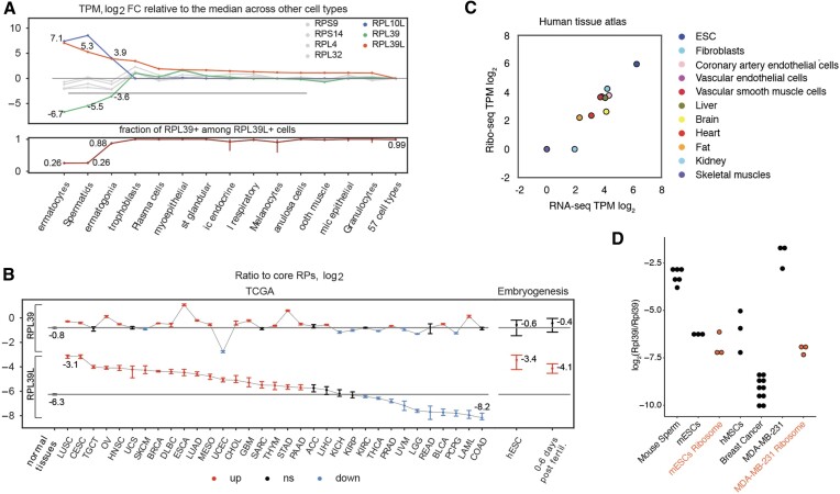
Aiming to determine the breadth of RPL39L expression across normal human cells, we examined the single-cell sequencing (scRNA-seq) data from The Human Protein Atlas (HPA (18), https://www.proteinatlas.org/download/rna_single_cell_type_tissue.tsv.zip). While most abundant in the cells of the male germ cell lineage, the RPL39L mRNA is also present in other cell types, such as the extravillous trophoblast, where the RPL39L level is ∼12-fold lower compared to spermatocytes (Figure 1A, top). In contrast, another germ cell-specific RP, RPL10L, is virtually absent outside of the male germ line (Figure 1A, top). For comparison, we also investigated the variation of mRNAs encoding core RPs (identified as described in Materials and methods, Supplementary Figure S1) across cell types, finding it to be much smaller relative to the RP paralogs (Figure 1A, top). The scRNA-seq data also gave us the opportunity to determine whether RPL39L replaces RPL39 in specific cell types or rather the two genes are co-expressed. In the HPA scRNA-seq read count data (https://www.proteinatlas.org/download/rna_single_cell_read_count.zip) all cells that contained RPL39L-derived reads also had RPL39-derived reads. The sole exception were male germ cells, most of which contained exclusively RPL39L reads. This suggests that some cell types have heterogeneous RPL39/RPL39L populations of ribosomes (Figure 1A, bottom panel). The relatively high expression level of RPL39L in trophoblast cells (Figure 1A. top) prompted us to further examine the dynamics of RPL39L/RPL39 expression in early human development relative to adult human tissues. As the overall abundance of ribosomes differs across cell types (2), we always examined the variation in RPL39L/RPL39 levels relative to the core RPs. Reanalyzing scRNA-seq data sets from pre-implantation embryos and embryonic stem cells (ESCs) (17) along with normal adult tissue samples in The Cancer Genome Atlas (https://www.cancer.gov/tcga/) we found that, relative to core RPs, RPL39 maintained the same level in embryonic and differentiated cells (Figure 1B), while the level of RPL39L was significantly higher in embryonic cells than in adult normal cells (Figure 1B). The RPL39L-to-core RP ratio also varied more than 30-fold across cancer types (Figure 1B), in many cancers reaching the values observed in embryonic cells. The ratio was highest in lung (LUSC) and cervix (CESC) tumors. In contrast, the RPL39-to-core RP ratio fluctuated much less across tumors (Ansari–Bradley test, P-value < 10−4) and relative to normal cells.
Figure 1.
RPL39L expression across cell types. (A) Top: HPA-provided normalized gene expression values (transcript-per-million, TPM) were used to identify the 14 cell types with highest RPL39L expression. The log2 fold change in each of these cell types relative to the median across all other 57 cell types in HPA is shown for RPL39L (orange), RPL39 (green), RPL10L (blue) genes and 4 core RPs (gray). Bottom: the proportion of RPL39L+ cells that also contained RPL39-derived reads in single cells of the types shown in the top panel. (B) Ratio of RPL39 and RPL39L to core RP expression (log2) in bulk RNA-seq samples of primary tumors from TCGA (https://www.cancer.gov/tcga/) (left panel) as well as human pre-implantation embryos and cultured embryonic stem cells (17) (right panel). See https://gdc.cancer.gov/resources-tcga-users/tcga-code-tables/tcga-study-abbreviations for TCGA cancer-type abbreviations. Black horizontal lines show the median values of these ratios in adult normal tissue samples from TCGA. Statistically significant (two-sided Wilcoxon test, Benjamini–Hochberg FDR < 0.05) positive and negative deviations from the medians are shown in orange and blue, respectively. For each category, the 95% confidence interval over all samples (for bulk data from TCGA) or all cells (for single-cell data) is shown. Categories for which the ratios were not significantly different from the median of the normal samples are shown in black. (C) Ribo-seq vs. RNA-seq level expression of RPL39L in samples from the human tissue atlas (extracted from the supplementary material of (63)). (D) Quantification of RPL39L/RPL39 protein ratio in various cellular systems (mouse sperm cells −6 samples, breast cancer cell line MDA-MB-231, bone marrow-derived mesenchymal stem cells (hMSC) and E14 mouse embryonic stem cells (mESC) −3 independent samples, breast cancer tissues −10 samples) using reference peptides. Similar quantification from purified ribosomes of E14 and MDA-MB-231 cells is also shown.
To determine whether the RPL39L mRNA is indeed translated into protein, we examined an extensive ribosome footprinting dataset from human primary cells and tissues (63). As shown in Figure 1C, we found the number of RPL39L-derived ribosome footprints to be largely proportional to the number of RPL39L-derived RNA-seq reads, indicating that the RPL39L mRNA does indeed undergo translation. Furthermore, human embryonic stem cells had relatively high expression of RPL39L, as we have seen in other datasets (Figure 1B). We also sought to measure the RPL39L protein in a few cellular systems. As RPL39L-specific antibodies are not available, we turned to mass spectrometry. Due to its high lysine/arginine content, the RPL39L protein is not reliably captured in standard proteomics analyses. Thus, we modified the sample preparation, acetylating the lysines in the proteome to direct the cleavage by trypsin to arginines only. We used heavy-labeled reference peptides to measure the RPL39L-to-RPL39 protein ratio in mature mouse sperm cells as positive control, and then in mouse embryonic stem cells, the human MDA-MB-231 breast cancer cell line, as well as human tissue samples, namely bone marrow-derived mesenchymal stem cells and breast cancer tissue. RPL39L was detectable at the protein level in these cell types, albeit at lower levels compared to mouse sperm (Figure 1D). To further answer the question of whether the protein is incorporated into ribosomes, we carried out the mass spectrometric analysis on ribosome populations purified by sucrose cushioning from E14 and MDA-MB-231 cell lines. The proportion of Rpl39l in the ribosomes of E14 mESC cells was similar to the proportion in the total lysate, while in the MDA-MB-231 cell line the proportion was much lower than in total lysate.
In human breast cancer tissue samples with heterogeneous cell type composition, RPL39L was also detectable, at lower abundance compared to pluripotent stem cells (Figure 1D). Our data thus provide conclusive mass spectrometric evidence of RPL39L protein expression not only in male germ cells, but also in pluripotent cells and cancer cell lines. The broader expression pattern of RPL39L compared to other germ cell-biased RP paralogs points to the relevance of RPL39/RPL39L ribosome heterogeneity beyond spermatogenesis.
KO of RPL39L impairs the pluripotency of E14 mESCs
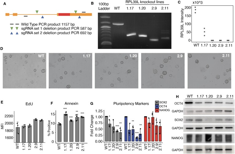
To determine the role of RPL39L in pluripotent cells we generated RPL39L knockout (KO) mESCs by CRISPR-mediated deletion of the coding region of this gene in the E14 mouse stem cell line. To minimize the chance of interfering with RPL39 we designed the sgRNAs to target non-coding regions of RPL39L, which are not shared with RPL39. To further exclude sgRNA-specific off-target effects, we designed two sets of guide RNAs that matched non-coding regions around the RPL39L CDS (Figure 2A), and for each sgRNA set we selected clones that originated from independent editing events (Supplementary Figure S2). We comprehensively analyzed two distinct clones for each sgRNA pair, in which the editing of the RPL39L locus was validated both by qPCR amplification with probes that flanked the edited region, and by measuring the protein expression with targeted proteomics (Figure 2B, C). All four clones (labeled as 1.17, 1.20, 2.9 and 2.11) exhibited homozygous deletions of the RPL39L locus, resulting from a spectrum of editing events including seemingly distinct deletions on the two chromosomes in clone 2.11 (Supplementary Figure S2). The RPL39L protein was undetectable in all of the edited clones except for 1.17, where some residual protein expression was detected.
Figure 2.
Characterization of RPL39L KO mESCs. (A) Schema of sgRNA design. Two distinct pairs of sgRNAs (in green and blue) were designed to target flanking regions of the RPL39L coding sequence (CDS, shown as a red box). Primers (black lines) from further upstream and downstream in the RPL39L locus were used for amplification, and the expected sizes of the PCR products are indicated for both the WT locus (1157 nts) and the edited loci (587 and 692 nts, respectively, for the sgRNA sets 1 and 2). (B) PCR products from the WT E14 cells and the 4 independent clones, 1.17 and 1.20 generated with sgRNA set #1 and2.9 and 2.11 generated with sgRNA set #2. (C) Expression of RPL39L in individual clones determined with targeted proteomics (n = 3 for each clone). (D) Representative bright field images of colonies from all analyzed clones. (E) Results of the ethynyldeoxyuridine (EdU, thymidine analog) incorporation assay. EdU-treated cells were fixed, permeabilized and the AF488 fluorophore was linked to the EdU in the replicated DNA by click-chemistry. The label intensity was measured by FACS (Mean fluorescence intensity, MFI). (F) Results of the annexin binding assay. Cells were incubated with AF488-conjugated Annexin V and counterstained with phycoerythrin (PE). The proportion of AF488+PE− (apoptotic cells) relative to the parent cell population was determined by FACS. (G) RT-qPCR of the pluripotency factors SOX2, OCT4 and NANOG. Values are 2−ΔΔCt, relative to RRM2 (internal reference) and to WT. (H) Representative Western blots of pluripotency markers SOX2, OCT4 and NANOG in the RPL39L KO lines and WT relative to GAPDH. In panels F and G, *, ** and *** correspond to P-values <0.05, <0.01 and < 0.001, respectively in the two tailed t-test comparing KO lines with WT.
The RPL39L KO cells were viable in culture, forming colonies that were similar in morphology to those formed by the WT mESCs (Figure 2D). Analysis of 5-ethynyl-2-deoxiuridine (EdU) incorporation during 2 h of treatment did not reveal significant differences between KO and WT cells, indicating that the RPL39L KO does not significantly impact proliferation in mESCs (Figure 2E). Annexin V staining showed only small, though statistically significant increase in the proportion of apoptotic cells relative to WT (Figure 2F). Despite no obvious changes in the colony morphology, the pluripotency markers exhibited significant variation; the Sox2 and Oct4 levels were reduced in KO compared to WT E14 lines, while Nanog showed less consistent reduction (Figure 2G). These differences were also apparent at the protein level (Figure 2H). Thus, RPL39L KO lines are viable, but have reduced expression of pluripotency markers relative to WT mESCs.
RPL39L KO mESCs exhibit differentiation defects
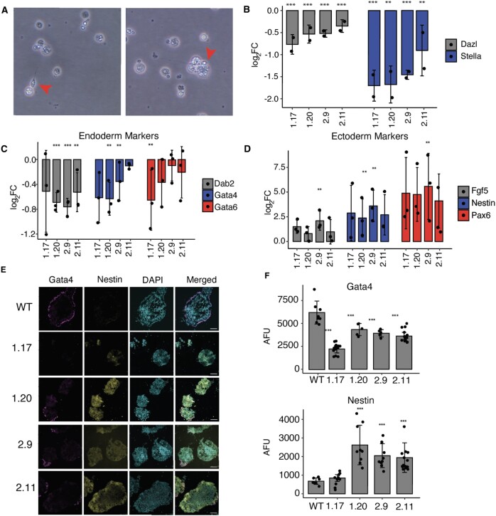
The perturbed expression of pluripotency markers in RPL39L KO cells prompted us to further investigate their ability to differentiate. Given the relevance of RPL39L for the spermatogenic lineage (11,64), we first subjected WT and RPL39L KO E14 lines to in vitro differentiation along this lineage using a previously described protocol (33). The emergence of spermatocyte-like cells in the WT E14 mESC culture demonstrated that the protocol works as expected (Figure 3A). In contrast, we did not identify any spermatocyte-like cells in the cultures of RPL39L KO lines, and the expression of germ-cell lineage markers Stella and Dazl were significantly reduced relative to differentiating WT cells (Figure 3B). These results show that RPL39L is necessary for spermatogenesis in vitro, consistent with previous observations in mice (11,64). To determine whether the KO of RPL39L impacts other developmental lineages as well, we carried out spontaneous differentiation of all our mESC lines in vitro, by culturing the cells in leukemia inhibiting factor (LIF)-free medium under non-adherent conditions (65). We then analyzed the expression of lineage markers both by qRT-PCR and by immunofluorescence-based quantification of protein levels in embryoid body sections (Figure 3C–F). The qRT-PCR revealed decreased expression of extraembryonic endoderm markers GATA6, GATA4and DAB2 in the organoids generated from the KO cells relative to those generated from WT cells (Figure 3C, E) and increased expression of ectodermal lineage markers NESTIN, FGF5 and PAX6 (Figure 3D, E). The protein level quantification from confocal images of NESTIN (ectoderm) and GATA4 (endoderm) validated the mRNA-level results (Figure 3F). Thus, the KO of RPL39L impacts spontaneous differentiation of mESCs, primarily towards the ectoderm and endoderm.
Figure 3.
RPL39L KO leads to differentiation defects in E14 mESCs. (A) Representative bright field images showing the spermatogenic differentiation of WT E14 cells. Red arrows indicate spermatocyte-like cells. (B) qRT-PCR assays of DAZL (late) and STELLA (early) sperm cell markers (33) (y-axis, log2 fold-change) in differentiating (4 days in RA-containing medium) populations of KO clones (x-axis) relative to WT. (C) qRT-PCR of extraembryonic endoderm markers (DAB2, GATA4, GATA6) (65) in RPL39L KO lines relative to WT. (D) Similar for FGF5, NESTIN, and PAX6 ectoderm markers (65). (E) Immunofluorescence staining of embryoid bodies subjected to spontaneous differentiation: GATA4 was used as endoderm marker, NESTIN as ectoderm marker and DAPI to delineate the nucleus. (F) Quantification of GATA4 and NESTIN expression in immunofluorescence images. AFU - arbitrary fluorescence units normalized to DAPI. In all panels, *, ** and *** correspond to P-values <0.05, <0.01 and < 0.001, respectively in the two tailed t-test comparing KO lines with WT.
RPL39L KO lines exhibit perturbed protein synthesis and ER stress
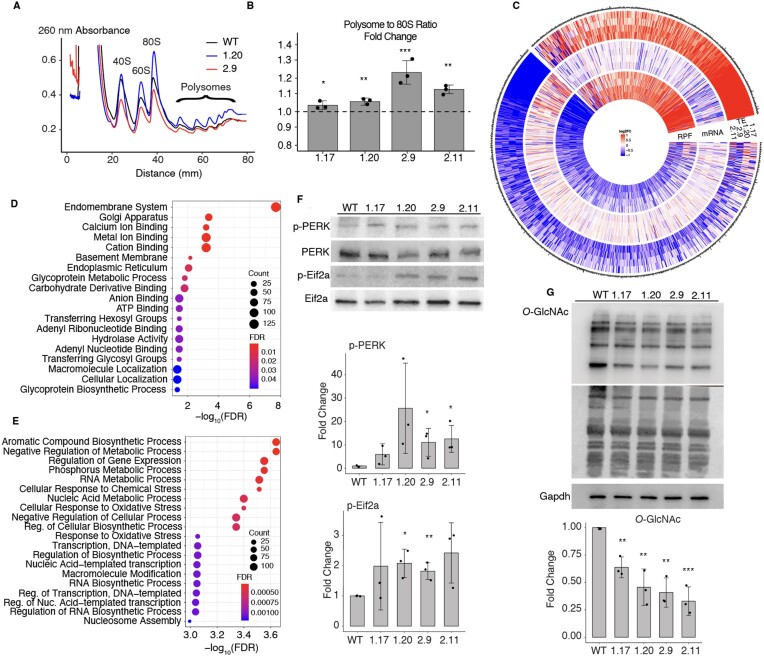
RPL39L being a ribosome component, to unravel the mechanisms underlying the observed functional defects, we evaluated the effect of RPL39L KO on global translation. We generated polysome profiles from all clones, and found that the KO of RPL39L leads to a small, but statistically significant increase in polysome-to-monosome ratio (Figure 4A, B). This indicates that RPL39L KO induces either a small increase in the rate of translation (66) or to an elongation defect (67) in mESCs.
Figure 4.
Impact of RPL39L KO on mRNA translation. (A) Example polysome profiles from the WT, 1.20 and 2.9 RPL39L KO E14 cell lines. (B) Ratio of the area under the profile corresponding to polysomes vs. monosomes (80S), in polysome profiles obtained from the KO clones. Fold-changes were calculated relative to the median ratio in the corresponding WT (dashed line at 1, n = 3 for all cell lines). (C) Log2 fold-changes in the translation efficiency (TE), mRNA level and the number of ribosome protected fragments (RPF) for specific genes, in mutant clones relative to WT E14 cells (n = 3 for each clone). Shown are all genes with a significant change in TE in at least one of the RPL39L KO clones. Values are capped at −1 and 1. (D, E) Gene Ontology analysis of mRNAs with reduced (D) and increased (E) TE in RPL39L KO clones. (F) Representative western blots and corresponding quantification (from n = 3 for each clone) of UPR markers PERK (phospho-Thr980) and EIF2A (phospho-Ser51). Intensities of phosphorylated proteins were normalized by the respective unphosphorylated forms and are relative to WT, for which the relative phosphorylation level was set to 1. (G) Representative western blot and corresponding quantification (from n = 3 for each clone) showing lower global O-GlcNAc modification of proteins in RPL39L KO lines when compared to the WT. Values are relative to GAPDH (loading control) and WT (level set to 1). In all panels, *, ** and *** correspond to P-values <0.05, <0.01 and < 0.001, respectively in the two tailed t-test comparing KO lines with WT.
To determine whether some transcripts are specifically impacted in translation by the RPL39L KO, we carried out ribosome footprinting (34), sequencing ribosome-protected mRNA fragments (RPFs) from both WT and RPL39L KO mESCs. The RPF data fulfilled expected quality criteria such as the vast majority of reads mapping to the coding regions of mRNAs and the 3 nucleotide periodicity of inferred P site locations (Supplementary Figure S3). We further sequenced the mRNAs from these cells and calculated the translation efficiency (TE) per mRNA as the ratio of the RPF and mRNA read density along the CDS (see Materials and methods). Focusing on mRNAs whose TE was significantly altered (P-value < 0.01 in ΔTE test, see Materials and methods) in at least one of the KO lines relative to the WT, we found that while the differences in RPFs and TE in KO clones relative to WT were generally small, the direction of change was highly consistent among specific classes of mRNAs (Figure 4C). Gene Ontology analysis showed that transcripts encoding components of the endomembrane system, including the Golgi apparatus and the endoplasmic reticulum (ER) experienced the strongest reduction in TE across the KO lines (Figure 4D). In contrast, we found a significantly increased TE for transcripts associated with the cellular response to chemical and oxidative stress (Figure 4E). Examples of increased and decreased RPF coverage of specific genes are shown in Supplementary Figure S3. These results suggest that the production of specific classes of proteins, associated with subcellular compartments such as the ER and Golgi apparatus, is impaired in RPL39L KO cells.
To further elucidate the relationship between the RPL39L KO and cellular stress, we measured the levels of both phosphorylated PKR-like ER kinase (PERK), a sensor of ER stress (68) and of its phosphorylation target, the eukaryotic initiation factor 2a (EIF2A) (69), which is responsible for regulation of many unfolded protein response (UPR)-associated stress-response genes. Western blotting showed that both of these markers were elevated in RPL39L KO compared to WT E14 cells (Figure 4F). We further used an O-GlcNAc antibody to label the glycosylated proteins from all our cell lines on a western blot. Integrating the chemiluminescence signal over the entire lanes corresponding to specific cell lines, we found reduced global signals in the samples from RPL39L KO cell lines compared to the WT E14 line (Figure 4G). Thus, ER stress markers are upregulated and protein glycosylation is impaired in RPL39L KO mESC lines relative to WT.
Increased degradation underlies the perturbed protein levels in RPL39L KO cell lines
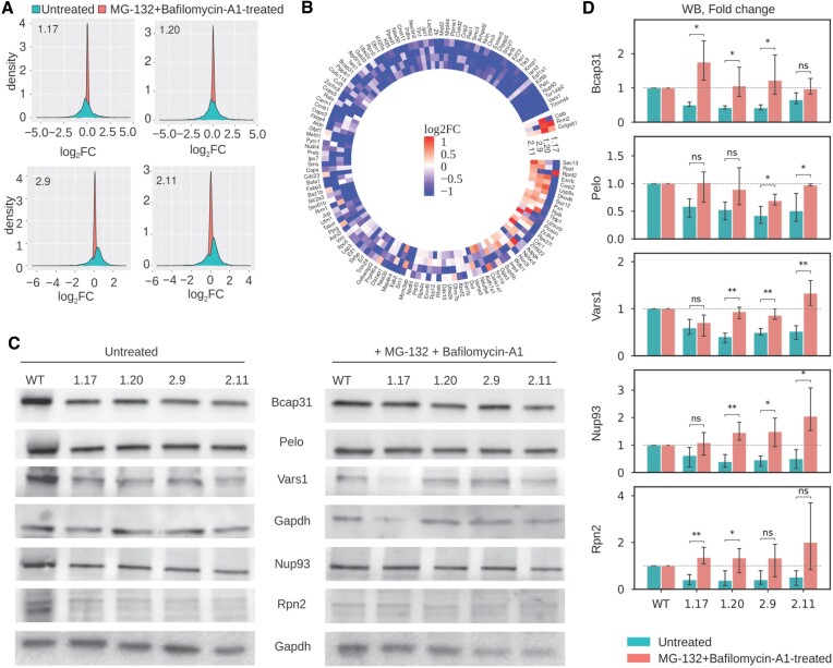
To learn more about the protein synthesis in RPL39L KO and WT cells, we measured the protein levels, both in steady-state and upon inhibition of proteasome and autophagy-dependent protein degradation (by treatment with MG132 and Bafilomycin A1, respectively), by shot-gun mass spectrometry. Overall, we identified 5026 proteins, 2992 of which in both treated and untreated conditions (Supplementary Figure S4). Strikingly, many proteins whose expression was perturbed by the RPL39L KO were restored to levels similar to those in WT by the inhibition of protein degradation, as indicated by the narrower distribution of log2 fold-changes of KO relative to WT cells in protease inhibitor-treated compared to untreated cells (Figure 5A). Along with the observed ER stress, this points to a reduced stability of proteins in RPL39L KO lines, which in turn suggests an increased production of defective proteins. This could be due to translation errors, e.g. amino acid misincorporation, or to defects in co-translational protein folding. The proteins whose reduced level in a KO clone relative to WT was restored by the treatment with protease inhibitors are shown in Figure 5B, which demonstrates their consistent behavior across KO clones. Western blotting further confirmed these results (Figure 5C, D). We further validated that the majority of the effect came from Bafilomycin A1, while MG132 contributed little, if at all, to rescuing the levels of proteins that are degraded in the RPL39L KO cells (Supplementary Figure S5). Gene Ontology analysis revealed enrichments in categories associated with the cytoskeleton and microtubules, indicating that the proteins stabilized by the protease inhibitor treatment are components of cellular membranes, contributing to cytoskeletal organization (Supplementary Figure S4). With a previously developed tool that specifically searches for amino acid substitutions in the measured peptides (49) we were unable to detect an enrichment of misincorporation in these proteins (Supplementary Figure S4). Thus, the most plausible explanation for the observed proteome changes is that RPL39L ribosomes facilitate the co-translational folding of specific proteins, leading to the production of defective proteins with compromised stability in the RPL39L KO clones.
Figure 5.
RPL39L KO clones exhibit enhanced degradation of specific classes of proteins. (A) Distribution of log2 fold changes in protein levels between untreated (blue) or MG-132 + Bafilomycin-A1-treated RPL39L KO and WT cells. Each panel corresponds to one KO clone. Three biological replicates for each condition were used to calculate average protein abundance levels and respective fold-changes relative to WT. (B) Heatmap of protein-level Δlog2 fold changes in KO cells relative to WT between untreated and treated cells. Included are all proteins with a significant downregulation in at least one of the untreated KO clones. (C) Representative western blot results showing the expression of a subset of proteins from (B) in untreated (left) and MG-132 + Bafilomycin-A1-treated cells (right). (D) Quantification of western blots as shown in (B), from three replicates for each protein and each condition.
Conformational differences between RPL39/RPL39L ribosome peptide exit tunnels
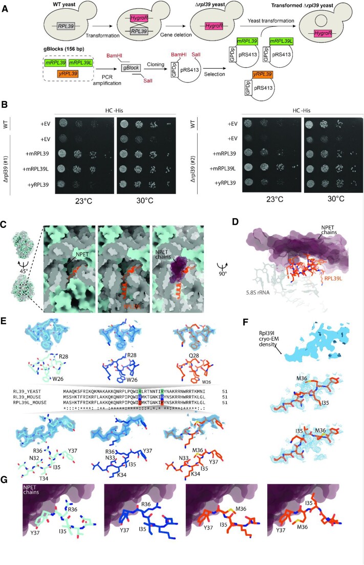
To understand how RPL39L could influence the dynamics of translation and the co-translational folding of proteins we sought to analyze RPL39L and RPL39-containing ribosomes by cryo-electron microscopy (cryo-EM). Mouse RPL39L has only three substitutions relative to RPL39: S2A, R28Q and R36M. Since these differences are subtle, structural investigations of RPL39/RPL39L-ribosomes necessitate high-resolution structures, which can only be obtained from homogeneous (RPL39 or RPL39L only) samples. Obtaining pure populations of RPL39L-containing ribosomes has been an unsolved challenge (11), which we have decided to overcome by expressing the mouse RPL39 and RPL39L in yeast RPL39 KO cell lines (Figure 6A). In yeast, just like in mammals, residues R28 and R36 of RPL39 face the lumen of the NPET and are in direct proximity to the nascent polypeptide chain.
Figure 6.
RPL39L introduces a hydrophobic patch in the NPET. (A) Scheme of yeast RPL39 KO and insertion of mouse RPL39 and mouse RPL39L. (B) The RPL39 KO causes growth defects in yeast upon environmental challenges like growth at 23 degrees Celsius in HC-His media. Expression of mouse RPL39 or RPL39L rescues these phenotypes. +EV are cells transformed with an empty vector as control. The experiments were carried out in two independent RPL39 knockout clones (#1 and #2). (C) RPL39L (cartoon representation, orange) shown in context of the large ribosomal subunit (refined atomic model of rpl39Δ-MmRpl39l; rRNAs are shown in gray, proteins are shown in pale blue surface representation). RPL39L is embedded inside the 60S subunit, adjacent to the exit of the NPET. Part of the 5.8S rRNA and ribosomal proteins have been removed for clarity on the center and right panel. Nascent protein chains and regulatory protein complexes pass the NPET in direct proximity to RPL39L as evident from aligned structures containing NPET-bound chains (nascent chains and regulatory proteins, PDB-IDs: 6M62, 7OBR, 7TM3, 7TUT, 7QWQ, 7QWS, shown as purple semi-transparent surfaces). (D) Side view of RPL39L (orange) and the surrounding 5.8S rRNA (gray) in direct contact with the NPET-facing region of RPL39L. The region containing Q28 and M36 is located directly adjacent to protein chains localized in the lumen of the NPET in structures containing nascent chains or tunnel-bound regulatory complexes (protein chain models shown as purple semi-transparent surfaces). (E) Comparison of the atomic model in the immediate surrounding of R/Q28 and R/M36 in WT yeast RPL39 (cyan), mouse RPL39 (dark blue), and mouse RPL39L (orange), shown in stick representation. The experimental cryo-EM density (semi-transparent surface, light blue) is shown superimposed on the refined atomic model. Maps around R28 are shown at a threshold of 6.5σ (yRPL39), 5.5σ (mRPL39), and 4.25σ (mRPL39L), while maps around R/M36 at a threshold of 5σ (yRPL39), 4σ (mRPL39), and 3.75σ (mRPL39L). Experimental cryo-EM density around M36 in mRPL39L is substantially weaker than the density observed either in yeast or mouse RPL39, due to increased conformational heterogeneity. (F) At a lower threshold, an alternative conformation is apparent in the experimental cryo-EM density (blue surface) of the region around M36 in mRPL39L, as RPL39L adopts two alternative conformations that differ substantially relative to the protein backbone and side chains. (G) In both WT yRPL39 (atomic model, stick representation, cyan) and mRPL39 (dark blue), the side chain of R36 faces the lumen of the NPET, potentially in direct contact with the protein chains inside the NPET (fitted chains of nascent protein and regulatory complexes, shown as purple semi-transparent surfaces). In mRPL39L, side chains of M36 and I35 hydrophobic residues are facing the NPET chains, forming a hydrophobic spot inside the NPET.
Thus, the replacement of R28 by Q28 and R36 by M36 in RPL39L, that is, of positively-charged residues by polar and hydrophobic residues, could perturb the co-translational folding or localization of proteins. The high degree of structural conservation of the 60S ribosomal subunit core in general, and of the region directly surrounding RPL39 in particular (mouse: RMSD = 0.52 Å, human: RMSD = 0.25 Å) further justifies the use of a heterologous system (70).
Increased rate of translation error reduces the growth of RPL39 KO (rpl39Δ) yeast strains at cold temperatures and sensitizes the cells to translation-interfering drugs like paromomycin (71) and azetidine-2-carboxylic acid (AZC) (72). We replicated these phenotypes in two distinct yeast RPL39 KO clones and further showed that they are rescued by the introduction of yRPL39 (rpl39Δ-ScRPL39 clones), mouse RPL39 (rpl39Δ-MmRPL39 clones) and mouse RPL39L (rpl39Δ-MmRPL39L clones) (Figure 6B and Supplementary Figure S6). This indicates that both mRPL39 and mRPL39L occupy the place of yRPL39 in the NPET and have a similar capacity to ensure the translation accuracy (71) and promote co-translational folding (72) in yeast. The results also suggest RPL39L shares the ancestral function of RPL39, though it may have acquired additional functions following its emergence in mammalian species.
Ribosomal particles from the WT yeast strain Sc60S, the yeast RPL39 KO strain rpl39Δ, yeast rescue strain rpl39Δ-ScRPL39 and the mouse RPL39/RPL39L-complemented RPL39 KO strains rpl39Δ-MmRPL39 and rpl39Δ-MmRPL39L were studied by single-particle cryo-EM (Supplementary Figures S7, S8). The structure of the rpl39Δ ribosome demonstrated the absence of RPL39 in the KO strain, with an otherwise structurally unaltered 60S subunit (overall RMSD = 0.308 Å), including the region surrounding RPL39 (RMSD = 0.38 Å) (Supplementary Figure S8). Expression of yRPL39 in a RPL39Δ background resulted in a correctly assembled 60S subunit that did not differ significantly structurally from the wild type 60S (RMSD = 0.192 Å), corroborating the feasibility of the complementation approach (Supplementary Figure S8). Mouse RPL39 and RPL39L both integrate into yeast 60S subunits when expressed in the rpl39Δ background (Supplementary Figure S8). Mouse RPL39, adopted a structure that was highly similar to that of RPL39 (RMSD = 0.482 Å) in the WT yeast ribosomes (Supplementary Figure S8). The structure of RPL39 in mouse (PDB: 6SWA, RMSD = 0.546 Å, (73)) and in human ribosomes (PDB: 7OW7 (74), RMSD = 0.451 Å) is also in agreement with the structure of rpl39Δ-MmRPL39, suggesting that our heterologous system adequately reflects the structure and conformation of mammalian RPL39 (Supplementary Figure S7). The comparison of rpl39Δ-MmRPL39 and rpl39Δ-MmRPL39L maps shows that the amino acids S2 in RPL39 and A2 in RPL39L are found at the same position, deeply embedded in rRNA 1490–1493, where they serve a structural role. The loop containing R28 and R36, exposed to the lumen of the NPET (Figure 6C–E), exhibits weaker cryo-EM density than the remainder of the protein in the map of rpl39Δ-MmRPL39. The exchange of R28 and R36 with glutamine and methionine, respectively, in RPL39L further increases the flexibility (Figure 6D-E) and enables the loop (residues 32–37) to adopt an alternative conformation where the Cɑ atom of I35 is displaced by 5.2 Å towards the exit of the NPET (Figure 6E-F). The positions of the basic R28 side chain in RPL39 and polar Q28 side chain in RPL39L are nearly identical. While residues M29, K30, T31, and G32 adopt the same conformation in RPL39 and RPL39L, the side chain of N33 has a slight displacement of Cγ: 1.87 Å. The side chain of K34, which is located in direct proximity to the phosphate backbone of A351 in RPL39, protrudes into the lumen of the tunnel in RPL39L. However, a partial occlusion of the tunnel appears unlikely as the side-chain cryo-EM density for K34 is very weak. The position of I35, which is facing towards the wall of the tunnel (A351 and A42) in RPL39, is occupied by M36 in RPL39L, leaving I35 oriented towards the lumen of the tunnel in RPL39L in the alternative conformation (Figure 6G). These observed structural changes do not substantially alter the overall electrostatic surface potential of the tunnel-exposed surface of RPL39/RPL39L, which is overwhelmingly positive in both cases. However, they do affect a narrow part of the ribosomal tunnel, traversed by the nascent chain (Figure 6D, E). Specifically, in RPL39L, Q28 and M36/I35 are found directly adjacent to each other at the location occupied by R28 and R36 in RPL39, introducing a clearly defined hydrophobic patch at the surface of the tunnel (Figure 6G). This may influence the translation speed, efficiency or the co-translational folding, either via direct interaction with the nascent chain (Figure 6G) or via interaction with translation regulation machinery such as the NAC complex, which was shown to bind to RPL39 (75).
Altered translation dynamics of RPL39L KO-destabilized proteins
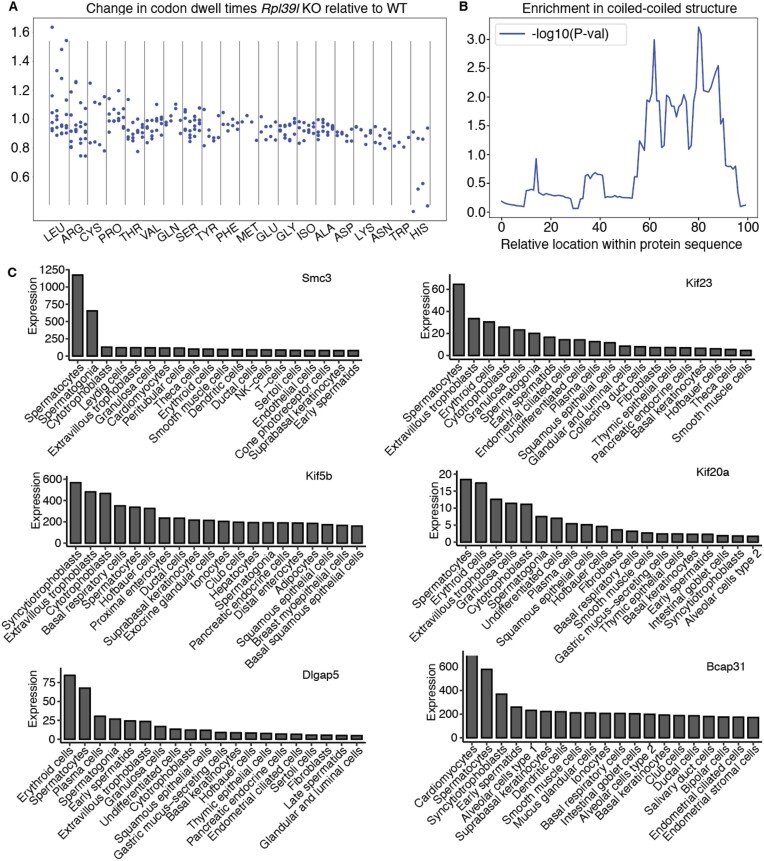
To determine how these conformational changes in the NPET may impact co-translational processes, we returned to the ribosome footprinting data, as such data were previously used to investigate multiple aspects of protein synthesis, including co-translational folding (reviewed in (76)). We estimated the change in ribosome dwell time on individual codons between WT and RPL39L KO clones as described before (77), constraining for the presence of a specific amino acid at a specific distance from the P site. We tested all 20 amino acids and all distances between 20 and 45 amino acids (Supplementary Figure S9). To identify consistent patterns of codon dwell time changes in independent Rpl39l KO clones, we calculated the cosine similarity scores between dwell time changes with respect to WT for pairs of clones and then the average across all pairwise comparisons. The highest average score, indicating the most consistent change in dwell times across Rpl39l KO clones, came from the amino acid leucine, when located at position +31 with respect to the P site. This is illustrated in Figure 7A, which shows that the presence of a leucine amino acid at position +31 leads to longer dwell times for leucine codons and shorter dwell time for the CAC codon of histidine. Histidine and leucine are amino acids that are enriched in helix-turn-helix, zinc finger, leucine zipper coiled-coil motifs, which occur in proteins of diverse functions (78–80). 7 of the 39 (18%) proteins that were consistently destabilized in the KO lines contained coiled-coils, which is ∼2-fold higher than the ∼8% estimated frequency of coiled-coil domain-containing proteins in the human proteome (81). We also estimated the proportion of amino acids involved in coiled-coiled domains as a function of their relative location in the sequence and then compared this fraction between proteins that were consistently destabilized in the KO lines and those that did not change. Mann–Whitney U test showed a significantly higher fraction of coiled-coil structure in the C-terminal half of presumed RPL39L targets compared to non-targets (Figure 7B). Interestingly, in the Human Proteome Atlas, 6 of the seven proteins (SMC3, KIF23, KIF5B, KIF20A, DLGAP5, BCAP31) have spermatocytes and/or trophoblast as the tissues of highest expression (Figure 7C). These are the tissues where RPL39L expression is also highest. Along with the ribosome profiling and protein stability data, these data suggest a role of RPL39L ribosomes in the co-translational folding of a subclass of proteins.
Figure 7.
Altered codon dwell times in RPL39l KO relative to WT clones. (A) Change in codon dwell time in the RPL39L KO clones relative to WT, conditioned on the presence of leucine at position +31 in the NPET. For each amino acid the 4 columns correspond to the 4 independent clones, shown always in the same order: 1_17, 1_20, 2_9 and 2_11. (B) Location-dependent P-values in the Mann–Whitney U test comparing the fractions of amino acids in coiled-coil structures within 100 length bins in presumed Rpl39l (39) targets and non-target proteins (4720). (C) Expression level (TPM) of mRNAs encoding proteins with coiled-coil domains that are destabilized in the RPL39L KO clones. Shown are values obtained by scRNA-seq of various cell types in the Human Protein Atlas. The gene names are indicated at the top of each panel and the cell types are labeled on the x-axis.
Discussion
mRNA translation is carried out by the ribosome, a highly conserved molecular machine dating back to the last universal common ancestor of cellular life (82). Although a ribosome can, in principle, translate any of the mRNAs expressed in a cell, the protein output varies among mRNAs, dependent on the efficiency of translation initiation, the speed of peptide chain elongation (83,84), and factors such as the total number of ribosomes in the cell (85–87). In recent years, a variety of factors have been found to modify the protein output of mRNAs at the level of ribosomes, among which ribosomal protein paralogs (reviewed in (88)). The human and mouse genomes encode around 20 RP paralogs, which have a more restricted distribution across tissues than canonical RPs (2). RPL39L is a recently evolved RP paralog (1) recently found to be critical to male fertility (11,64). The RPL39L mRNA is also expressed outside of the male germ line, in normal and cancer cells (2,12,13), where its role is still unknown. Furthermore, while RPL39L has been implicated in the folding of long-lived sperm proteins (11,64), how this is achieved is unclear.
To answer these questions, we generated RPL39L mESC KO lines by CRISPR-mediated genome editing. ESCs naturally express RPL39L, as we have found in analyses of bulk and scRNA-seq datasets. To confirm RPL39L expression at the protein level, we developed a mass spectrometric-based approach wherein lysine residues are acetylated proteome-wide to direct the tryptic cleavage to arginine residues. With its high content of lysines and arginines (31% of 51 amino acids), RPL39L is almost completely digested by trypsin during standard sample preparation for mass spectrometry, leaving only incompletely cleaved peptides amenable for detection. This may explain why prior mass spectrometric analyses have not detected this protein in tissues with relatively low frequency of RPL39L-expressing cells. If RPL39 and RPL39L carry their functions as part of ribosomes, as has been suggested before (11,89), the RPL39L/RPL39 ratios that we estimated imply that ∼1% of the ribosomes in pluripotent cells contain RPL39L. Indeed, mass spectrometry data of ribosomes purified by sucrose cushioning corroborates this result. Moreover, single cells that contain RPL39L reads also contain RPL39 reads, which indicates that pluripotent cells have heterogeneous RPL39/RPL39L ribosome populations. The relatively high expression of RPL39L mRNA in extravillous trophoblast, breast glandular and pancreatic endocrine cells from the Human Protein Atlas, as well as multiple cancer types, suggested that RPL39L may support protein synthesis in the context of secretory processes.
By morphological and molecular characterization of KO cell lines, we demonstrated that RPL39L contributes to mESC pluripotency and differentiation along multiple lineages. Like RPL10L, RPL39L is a recently evolved autosomal chromosome-encoded paralog of an X chromosome-encoded RP. These paralogs are thought to take part in the formation of testis-specific ribosomes, necessary for spermatogenesis (89). Indeed, RPL10L has been shown to compensate for the reduced level of RPL10 during meiotic sex chromosome inactivation (10), while mice deficient in RPL39L exhibit spermatogenesis defects (11,64). We found that RPL39L KO mESCs are not only impaired in the ability to differentiate into sperm cells but also show defects in spontaneous differentiation. The expression of ectoderm markers was increased and of endoderm markers decreased in embryoid bodies derived from RPL39L KO mESCs compared to those derived from WT cells. Whether these defects that we observed in vitro can also be detected in the early mouse development has not been investigated (11,64).
To evaluate the impact of RPL39L on translation, we carried out ribosome profiling (34). Consistent with previous studies (11,64), we found a small upregulation in the polysome-to-monosome ratio in the KO compared to WT clones, which indicates an increased protein synthesis rate. At the level of individual mRNAs, the changes in translation efficiency were very consistent across KO clones and occurred in mRNAs with specific functions. ER and Golgi-associated membrane proteins were generally less translated, while stress response and oxidative damage-related proteins were more translated in the RPL39L KO compared to WT mESCs. The increased phosphorylation of PERK and EIF2A further indicated that the stress was due to the ER-associated unfolded protein response (UPR) (90), consistent with an apparent increase in protein degradation that we also consistently found across all RPL39L KO clones. Pluripotent cells have higher resistance to ER stress than differentiated cells. However, the normal differentiation along the endodermal and ectodermal lineages requires UPR to be maintained at a specific level (91), which may explain why differentiation is perturbed as the mESCs experienced increased ER stress upon RPL39L KO.
We demonstrated that the reduced protein levels in RPL39L KO relative to WT mESCs were restored by the inhibition of autophagy, showing that the protein stability is impaired in RPL39L KO cells, as also demonstrated by previous works (11). Some of the destabilized proteins have a testis bias of expression (according to the mRNA level data in the HPA), but the set more generally included proteins that are involved in cytoskeleton organization, cellular localization (specifically to membranes) and polarization. As we did not find an increased frequency of amino acid misincorporation in the KO clones, we investigated whether the decreased protein stability is due to alterations in other co-translational processes such as folding. RPL39 is known to interact with transmembrane alpha helices that fold in the NPET, but not with the alpha helices of secretory proteins (92). To elucidate the origin of these interactions, we turned to structural analyses. Obtaining pure populations of RPL39L ribosomes is very challenging and has not been achieved so far. Thus, to determine how RPL39/RPL39L impact the NPET structure we overexpressed the mouse RPL39 and RPL39L in RPL39 KO yeast strains. The RPL39 KO yeast lines are sensitive to cold, paromomycin and AZT, phenotypes that have been described before (71,72). These phenotypes are rescued by both mouse RPL39 and mouse RPL39L, which indicates that RPL39L can perform the basic functions of RPL39. Analysis of single ribosome particles from these RPL39/RPL39L-expressing strains revealed that the main structural difference is a conformational change that involves the conserved residue I35, and leads to the appearance of a hydrophobic patch in the vestibular region of NPET, in the position where RPL39-containing ribosomes expose a positive charge. The loop containing Q28 and M36 further shows a higher flexibility compared to that provided by RPL39. Notably, we did not find electron densities supporting an occlusion of the tunnel region by RPL39L as proposed by a previous study (11). In fact, we did not find such evidence upon re-examining the published data either. Thus, it seems unlikely that RPL39L promotes the folding of specific proteins by occluding the NPET. What other mechanism could be responsible? Early studies hypothesized that a hydrophobic patch near the vestibular region of the NPET nucleates the co-translational folding of alpha helices (15). While the first structures of the 60S subunit of an eubacterial ribosome (93) did not identify such a patch (94), in prokaryotes, the uL23 RP, located at the vestibular region of the NPET recruits the trigger factor a (TFa) (95) to provide the hydrophobic surface for folding of cytosolic hydrophobic proteins (96). In eukaryotes, RPL39 replaces a loop of uL23 in the lower part of the tunnel, making it considerably narrower (97), which is thought to promote the entropic stabilization of alpha helices (98), and probably reducing the need for the hydrophobic patch to promote nascent protein folding. However, a narrower tunnel also leads to a reduced speed and increased accuracy of translation, both of which are impacted by the deletion of RPL39 (71). It is remarkable that the distinguishing feature of RPL39L ribosomes is the re-emergence of the hydrophobic patch, provided by RPL39L instead of the TFa chaperone, as chaperone function has diversified during the evolution of eukaryotes (99).
Although our study was done in mESCs, we found that the RPL39L-sensitive proteins have a testis bias of expression. To further understand the properties of these RPL39L-sensitive proteins, we further analyzed the dynamics of translation by calculating ribosome dwell time on individual codons and on codons encoding individual amino acids. We further carried out this analysis conditioning the calculation on the presence of specific amino acids at specific distances upstream of the P-site of the ribosome. We found between clones-consistent alterations in codon dwell time relative to WT particularly when leucine was present at position + 31. In this case (though also more generally) dwell times increased for leucine codons and decreased for the CAC codon of histidine in RPL39L KO cells compared to WT. These amino acids occur in helix-turn-helix, zinc finger, and leucine zipper coiled coil motifs (100), which are found in amphiphilic alpha helices (101,102). The mRNA level data from HPA further shows that proteins with coiled-coil domains that are destabilized in the RPL39L KO mESCs exhibit an expression pattern surprisingly similar to that of RPL39L, in that they have highest mRNA-level expression in the spermatogenic lineage and/or trophoblast. Although amphiphilic alpha helices can fold in RPL39-containing ribosomes, the presence of a hydrophobic patch and better hydrophilic/hydrophobic partitioning may allow a more reliable folding of such domains, important when the burden of protein production is high, as in secretory cells (glandular, endocrine etc.) or cells that undergo large changes in polarity (developing sperm cells, ES cells etc.) (103,104).
Our work elucidates the function of a specialized ribosome, defined by RPL39L and present in sperm cells, but also other cell types such as mESCs. However, additional questions remain to be addressed in the future. First, the implications of RPL39L KO on the in vivo animal development are insufficiently understood. As already mentioned, RPL39L-deficient mice obtained via CRISPR/Cas9 genome editing (11,64) were not investigated beyond the spermatogenesis defect. At the molecular level, it will be interesting to investigate further how ES cells and cancer cells fine-tune their translation output and the quality of synthesized proteins using modular ribosome components.
Supplementary Material
Acknowledgements
We are grateful to Dr Constance Ciaudo for suggestions regarding mESC differentiation, to Dr Arnaud Schieberich for the hBM-MSC cells, Dr Frederic Schmitt for the mouse samples, Dr. Sebastian Hiller and Dr Nenad Ban for insightful discussions on the ribosome structure, and Dr Vinay Tergaonkar for suggestions on the project. We thank the Imaging Core Facility at Biozentrum (IMCF), especially Laurent Guerard, Dr Sara Roig and Dr Kai Schleicher for help with the image acquisition and data analysis, and the sciCORE team for supporting the computational infrastructure. We would also like to thank Stella Stefanova and Janine Boegli of Biozentrum FACS core facility for help with the FACS data acquisition, and Philippe Demougin and Dr Christian Beisel from the genomics facility Basel for help with RNA sequencing. Cryo-EM data were collected in the Scientific Center for Optical and Electron Microscopy (ScopeM). We thank Miroslav Peterek (ScopeM) for support and Pavel Afanasyev (CEMK) for helpful discussions. This work was supported in part by the SNF grant #51NF40_141735 for the NCCR project ‘RNA & Disease’ in which the Zavolan group participated. The results shown here are in part based upon data generated by the TCGA Research Network: https://www.cancer.gov/tcga. Graphical abstract was created with Biorender.com.
Contributor Information
Arka Banerjee, Biozentrum, University of Basel, Basel, Switzerland.
Meric Ataman, Biozentrum, University of Basel, Basel, Switzerland.
Maciej Jerzy Smialek, Biozentrum, University of Basel, Basel, Switzerland; Institute of Human Genetics, Polish Academy of Sciences, Poznan, Poland.
Debdatto Mookherjee, Biozentrum, University of Basel, Basel, Switzerland.
Julius Rabl, Cryo-EM Knowledge Hub (CEMK), ETH Zürich, Switzerland.
Aleksei Mironov, Biozentrum, University of Basel, Basel, Switzerland.
Lea Mues, Biozentrum, University of Basel, Basel, Switzerland.
Ludovic Enkler, Biozentrum, University of Basel, Basel, Switzerland; University of Strasbourg, UMR7156 GMGM, Strasbourg, France.
Mairene Coto-Llerena, Institute of Medical Genetics and Pathology, University Hospital Basel, University of Basel, Switzerland.
Alexander Schmidt, Biozentrum, University of Basel, Basel, Switzerland.
Daniel Boehringer, Cryo-EM Knowledge Hub (CEMK), ETH Zürich, Switzerland.
Salvatore Piscuoglio, Institute of Medical Genetics and Pathology, University Hospital Basel, University of Basel, Switzerland; IRCCS Humanitas Research Hospital, Rozzano, Milan, Italy.
Anne Spang, Biozentrum, University of Basel, Basel, Switzerland.
Nitish Mittal, Biozentrum, University of Basel, Basel, Switzerland.
Mihaela Zavolan, Biozentrum, University of Basel, Basel, Switzerland.
Data availability
Sequencing data has been deposited to the NCBI BioProject database (https://www.ncbi.nlm.nih.gov/bioproject/), under accession PRJNA951511, and the mass spectrometry to the MassIVE repository (https://massive.ucsd.edu/ProteoSAFe/static/massive.jsp), accession number MSV000091698. Processing scripts are available from the zenodo repository, with DOI 10.5281/zenodo.7794007. Cryo-EM data has been deposited: Mouse RPL39L integrated into the yeast 60S ribosomal subunit (PDB ID 8PFR) and EMDB entry ID EMD-17653; Mouse RPL39 integrated into the yeast 60S ribosomal subunit (PDB ID 8P8N) and EMDB entry ID EMD-17550; Yeast 60S ribosomal subunit, RPL39 deletion (PDB ID 8P8M) and EMDB entry ID EMD-17549; Yeast 60S ribosomal subunit (PDB ID 8P8U) and EMDB entry ID EMD-17552.
Supplementary data
Supplementary Data are available at NAR Online.
Funding
Schweizerischer Nationalfonds zur Förderung der Wissenschaftlichen Forschung [51NF40_141735]. Funding for open access charge: Schweizerischer Nationalfonds zur Förderung der Wissenschaftlichen Forschung.
Conflict of interest statement. None declared.
References
- 1. Wong Q.W.-L., Li J., Ng S.R., Lim S.G., Yang H., Vardy L.A.. RPL39L is an example of a recently evolved ribosomal protein paralog that shows highly specific tissue expression patterns and is upregulated in ESCs and HCC tumors. RNA Biol. 2014; 11:33–41. [DOI] [PMC free article] [PubMed] [Google Scholar]
- 2. Guimaraes J.C., Zavolan M.. Patterns of ribosomal protein expression specify normal and malignant human cells. Genome Biol. 2016; 17:236. [DOI] [PMC free article] [PubMed] [Google Scholar]
- 3. Xiong X., Zhao Y., He H., Sun Y.. Ribosomal protein S27-like and S27 interplay with p53-MDM2 axis as a target, a substrate and a regulator. Oncogene. 2011; 30:1798–1811. [DOI] [PMC free article] [PubMed] [Google Scholar]
- 4. Zhang Y., O’Leary M.N., Peri S., Wang M., Zha J., Melov S., Kappes D.J., Feng Q., Rhodes J., Amieux P.S.et al.. Ribosomal proteins Rpl22 and Rpl22l1 control morphogenesis by regulating pre-mRNA splicing. Cell Rep. 2017; 18:545–556. [DOI] [PMC free article] [PubMed] [Google Scholar]
- 5. Chaillou T., Zhang X., McCarthy J.J.. Expression of muscle-specific ribosomal protein L3-like impairs myotube growth. J. Cell. Physiol. 2016; 231:1894–1902. [DOI] [PMC free article] [PubMed] [Google Scholar]
- 6. Shiraishi C., Matsumoto A., Ichihara K., Yamamoto T., Yokoyama T., Mizoo T., Hatano A., Matsumoto M., Tanaka Y., Matsuura-Suzuki E.et al.. RPL3L-containing ribosomes determine translation elongation dynamics required for cardiac function. Nat. Commun. 2023; 14:2131. [DOI] [PMC free article] [PubMed] [Google Scholar]
- 7. Milenkovic I., Santos Vieira H.G., Lucas M.C., Ruiz-Orera J., Patone G., Kesteven S., Wu J., Feneley M., Espadas G., Sabidó E.et al.. Dynamic interplay between RPL3- and RPL3L-containing ribosomes modulates mitochondrial activity in the mammalian heart. Nucleic Acids Res. 2023; 51:5301–5324. [DOI] [PMC free article] [PubMed] [Google Scholar]
- 8. Uechi T., Maeda N., Tanaka T., Kenmochi N.. Functional second genes generated by retrotransposition of the X-linked ribosomal protein genes. Nucleic Acids Res. 2002; 30:5369–5375. [DOI] [PMC free article] [PubMed] [Google Scholar]
- 9. Sugihara Y., Sadohara E., Yonezawa K., Kugo M., Oshima K., Matsuda T., Nadano D.. Identification and expression of an autosomal paralogue of ribosomal protein S4, X-linked, in mice: potential involvement of testis-specific ribosomal proteins in translation and spermatogenesis. Gene. 2013; 521:91–99. [DOI] [PubMed] [Google Scholar]
- 10. Jiang L., Li T., Zhang X., Zhang B., Yu C., Li Y., Fan S., Jiang X., Khan T., Hao Q.et al.. RPL10L Is required for male meiotic division by compensating for RPL10 during meiotic sex chromosome inactivation in mice. Curr. Biol. 2017; 27:1498–1505. [DOI] [PubMed] [Google Scholar]
- 11. Li H., Huo Y., He X., Yao L., Zhang H., Cui Y., Xiao H., Xie W., Zhang D., Wang Y.et al.. A male germ-cell-specific ribosome controls male fertility. Nature. 2022; 612:725–731. [DOI] [PubMed] [Google Scholar]
- 12. Rohozinski J., Anderson M.L., Broaddus R.E., Edwards C.L., Bishop C.E.. Spermatogenesis associated retrogenes are expressed in the human ovary and ovarian cancers. PLoS One. 2009; 4:e5064. [DOI] [PMC free article] [PubMed] [Google Scholar]
- 13. Yan P., Yang X., Wang J., Wang S., Ren H.. A novel CpG island methylation panel predicts survival in lung adenocarcinomas. Oncol. Lett. 2019; 18:1011–1022. [DOI] [PMC free article] [PubMed] [Google Scholar]
- 14. PCAWG Transcriptome Core Group Calabrese C., Davidson N.R., Demircioğlu D., Fonseca N.A., He Y., Kahles A., Lehmann K.-V., Liu F., Shiraishi Y.et al.. Genomic basis for RNA alterations in cancer. Nature. 2020; 578:129–136. [DOI] [PMC free article] [PubMed] [Google Scholar]
- 15. Liao S., Lin J., Do H., Johnson A.E.. Both lumenal and cytosolic gating of the aqueous ER translocon pore are regulated from inside the ribosome during membrane protein integration. Cell. 1997; 90:31–41. [DOI] [PubMed] [Google Scholar]
- 16. Dobin A., Davis C.A., Schlesinger F., Drenkow J., Zaleski C., Jha S., Batut P., Chaisson M., Gingeras T.R.. STAR: ultrafast universal RNA-seq aligner. Bioinformatics. 2013; 29:15–21. [DOI] [PMC free article] [PubMed] [Google Scholar]
- 17. Yan L., Yang M., Guo H., Yang L., Wu J., Li R., Liu P., Lian Y., Zheng X., Yan J.et al.. Single-cell RNA-seq profiling of human preimplantation embryos and embryonic stem cells. Nat. Struct. Mol. Biol. 2013; 20:1131–1139. [DOI] [PubMed] [Google Scholar]
- 18. Uhlén M., Fagerberg L., Hallström B.M., Lindskog C., Oksvold P., Mardinoglu A., Sivertsson Å., Kampf C., Sjöstedt E., Asplund A.et al.. Proteomics. Tissue-based map of the human proteome. Science. 2015; 347:1260419. [DOI] [PubMed] [Google Scholar]
- 19. Kater L., Thoms M., Barrio-Garcia C., Cheng J., Ismail S., Ahmed Y.L., Bange G., Kressler D., Berninghausen O., Sinning I.et al.. Visualizing the assembly pathway of nucleolar pre-60S ribosomes. Cell. 2017; 171:1599–1610. [DOI] [PMC free article] [PubMed] [Google Scholar]
- 20. de la Cruz J., Karbstein K., Woolford J.L. Jr. Functions of ribosomal proteins in assembly of eukaryotic ribosomes in vivo. Annu. Rev. Biochem. 2015; 84:93–129. [DOI] [PMC free article] [PubMed] [Google Scholar]
- 21. Razi A., Ortega J.. Ribosomal proteins: their role in the assembly, structure and function of the ribosome. eLS. 2017; John Wiley & Sons, Ltd; 10.1002/9780470015902.a0000535.pub2. [DOI] [Google Scholar]
- 22. Hu Y., Flockhart I., Vinayagam A., Bergwitz C., Berger B., Perrimon N., Mohr S.E.. An integrative approach to ortholog prediction for disease-focused and other functional studies. BMC Bioinf. 2011; 12:357. [DOI] [PMC free article] [PubMed] [Google Scholar]
- 23. Pacini C., Dempster J.M., Boyle I., Gonçalves E., Najgebauer H., Karakoc E., van der Meer D., Barthorpe A., Lightfoot H., Jaaks P.et al.. Integrated cross-study datasets of genetic dependencies in cancer. Nat. Commun. 2021; 12:1661. [DOI] [PMC free article] [PubMed] [Google Scholar]
- 24. McFarland J.M., Ho Z.V., Kugener G., Dempster J.M., Montgomery P.G., Bryan J.G., Krill-Burger J.M., Green T.M., Vazquez F., Boehm J.S.et al.. Improved estimation of cancer dependencies from large-scale RNAi screens using model-based normalization and data integration. Nat. Commun. 2018; 9:4610. [DOI] [PMC free article] [PubMed] [Google Scholar]
- 25. Frankish A., Diekhans M., Jungreis I., Lagarde J., Loveland J.E., Mudge J.M., Sisu C., Wright J.C., Armstrong J., Barnes I.et al.. Gencode 2021. Nucleic Acids Res. 2021; 49:D916–D923. [DOI] [PMC free article] [PubMed] [Google Scholar]
- 26. Graubert A., Aguet F., Ravi A., Ardlie K.G., Getz G.. RNA-SeQC 2: efficient RNA-seq quality control and quantification for large cohorts. Bioinformatics. 2021; 37:3048–3050. [DOI] [PMC free article] [PubMed] [Google Scholar]
- 27. Zhang Z., Harrison P., Gerstein M.. Identification and analysis of over 2000 ribosomal protein pseudogenes in the human genome. Genome Res. 2002; 12:1466–1482. [DOI] [PMC free article] [PubMed] [Google Scholar]
- 28. Pertea M., Pertea G.M., Antonescu C.M., Chang T.-C., Mendell J.T., Salzberg S.L.. StringTie enables improved reconstruction of a transcriptome from RNA-seq reads. Nat. Biotechnol. 2015; 33:290–295. [DOI] [PMC free article] [PubMed] [Google Scholar]
- 29. Peterson A.C., Russell J.D., Bailey D.J., Westphall M.S., Coon J.J.. Parallel reaction monitoring for high resolution and high mass accuracy quantitative, targeted proteomics. Mol. Cell. Proteomics. 2012; 11:1475–1488. [DOI] [PMC free article] [PubMed] [Google Scholar]
- 30. Gallien S., Duriez E., Crone C., Kellmann M., Moehring T., Domon B.. Targeted proteomic quantification on quadrupole-orbitrap mass spectrometer. Mol. Cell. Proteomics. 2012; 11:1709–1723. [DOI] [PMC free article] [PubMed] [Google Scholar]
- 31. MacLean B., Tomazela D.M., Shulman N., Chambers M., Finney G.L., Frewen B., Kern R., Tabb D.L., Liebler D.C., MacCoss M.J.. Skyline: an open source document editor for creating and analyzing targeted proteomics experiments. Bioinformatics. 2010; 26:966–968. [DOI] [PMC free article] [PubMed] [Google Scholar]
- 32. Kurosawa H. Methods for inducing embryoid body formation: in vitro differentiation system of embryonic stem cells. J. Biosci. Bioeng. 2007; 103:389–398. [DOI] [PubMed] [Google Scholar]
- 33. Kerkis A., Fonseca S.A.S., Serafim R.C., Lavagnolli T.M.C., Abdelmassih S., Abdelmassih R., Kerkis I.. In vitro differentiation of male mouse embryonic stem cells into both presumptive sperm cells and oocytes. Cloning Stem Cells. 2007; 9:535–548. [DOI] [PubMed] [Google Scholar]
- 34. Ingolia N.T., Brar G.A., Rouskin S., McGeachy A.M., Weissman J.S.. The ribosome profiling strategy for monitoring translation in vivo by deep sequencing of ribosome-protected mRNA fragments. Nat. Protoc. 2012; 7:1534–1550. [DOI] [PMC free article] [PubMed] [Google Scholar]
- 35. Hornstein N., Torres D., Das Sharma S., Tang G., Canoll P., Sims P.A.. Ligation-free ribosome profiling of cell type-specific translation in the brain. Genome Biol. 2016; 17:149. [DOI] [PMC free article] [PubMed] [Google Scholar]
- 36. Hoffmann S., Otto C., Kurtz S., Sharma C.M., Khaitovich P., Vogel J., Stadler P.F., Hackermüller J.. Fast mapping of short sequences with mismatches, insertions and deletions using index structures. PLoS Comput. Biol. 2009; 5:e1000502. [DOI] [PMC free article] [PubMed] [Google Scholar]
- 37. Katsantoni M., Gypas F., Herrmann C.J., Burri D., Bak M., Iborra P., Agarwal K., Ataman M., Börsch A., Zavolan M.et al.. ZARP: an automated workflow for processing of RNA-seq data. F1000Research. (2024); 13:533. [Google Scholar]
- 38. Bray N.L., Pimentel H., Melsted P., Pachter L.. Near-optimal probabilistic RNA-seq quantification. Nat. Biotechnol. 2016; 34:525–527. [DOI] [PubMed] [Google Scholar]
- 39. Love M.I., Huber W., Anders S.. Moderated estimation of fold change and dispersion for RNA-seq data with DESeq2. Genome Biol. 2014; 15:550. [DOI] [PMC free article] [PubMed] [Google Scholar]
- 40. Chothani S., Adami E., Ouyang J.F., Viswanathan S., Hubner N., Cook S.A., Schafer S., Rackham O.J.L.. deltaTE: detection of translationally regulated genes by integrative analysis of ribo-seq and RNA-seq data. Curr. Protoc. Mol. Biol. 2019; 129:e108. [DOI] [PMC free article] [PubMed] [Google Scholar]
- 41. Liao Y., Smyth G.K., Shi W.. The R package Rsubread is easier, faster, cheaper and better for alignment and quantification of RNA sequencing reads. Nucleic Acids Res. 2019; 47:e47. [DOI] [PMC free article] [PubMed] [Google Scholar]
- 42. Yu G., Wang L.-G., Han Y., He Q.-Y.. clusterProfiler: an R package for comparing biological themes among gene clusters. Omics. 2012; 16:284–287. [DOI] [PMC free article] [PubMed] [Google Scholar]
- 43. Gu Z., Eils R., Schlesner M.. Complex heatmaps reveal patterns and correlations in multidimensional genomic data. Bioinformatics. 2016; 32:2847–2849. [DOI] [PubMed] [Google Scholar]
- 44. Gu Z., Gu L., Eils R., Schlesner M., Brors B.. circlize implements and enhances circular visualization in R. Bioinformatics. 2014; 30:2811–2812. [DOI] [PubMed] [Google Scholar]
- 45. Heberle H., Meirelles G.V., da Silva F.R., Telles G.P., Minghim R.. InteractiVenn: a web-based tool for the analysis of sets through Venn diagrams. BMC Bioinf. 2015; 16:169. [DOI] [PMC free article] [PubMed] [Google Scholar]
- 46. UniProt Consortium UniProt: the Universal Protein knowledgebase in 2023. Nucleic Acids Res. 2023; 51:D523–D531. [DOI] [PMC free article] [PubMed] [Google Scholar]
- 47. Hastie T., Tibshirani R., Narasimhan B., Chu G. 2017; Impute bioconductor. [DOI] [PMC free article] [PubMed]
- 48. Ritchie M.E., Phipson B., Wu D., Hu Y., Law C.W., Shi W., Smyth G.K.. limma powers differential expression analyses for RNA-sequencing and microarray studies. Nucleic Acids Res. 2015; 43:e47. [DOI] [PMC free article] [PubMed] [Google Scholar]
- 49. Mordret E., Dahan O., Asraf O., Rak R., Yehonadav A., Barnabas G.D., Cox J., Geiger T., Lindner A.B., Pilpel Y.. Systematic detection of amino acid substitutions in proteomes reveals mechanistic basis of ribosome errors and selection for translation fidelity. Mol. Cell. 2019; 75:427–441. [DOI] [PubMed] [Google Scholar]
- 50. Tyanova S., Temu T., Cox J.. The MaxQuant computational platform for mass spectrometry-based shotgun proteomics. Nat. Protoc. 2016; 11:2301–2319. [DOI] [PubMed] [Google Scholar]
- 51. Schindelin J., Arganda-Carreras I., Frise E., Kaynig V., Longair M., Pietzsch T., Preibisch S., Rueden C., Saalfeld S., Schmid B.et al.. Fiji: an open-source platform for biological-image analysis. Nat. Methods. 2012; 9:676–682. [DOI] [PMC free article] [PubMed] [Google Scholar]
- 52. Burel J.-M., Besson S., Blackburn C., Carroll M., Ferguson R.K., Flynn H., Gillen K., Leigh R., Li S., Lindner D.et al.. Publishing and sharing multi-dimensional image data with OMERO. Mamm. Genome. 2015; 26:441–447. [DOI] [PMC free article] [PubMed] [Google Scholar]
- 53. Ershov D., Phan M.-S., Pylvänäinen J.W., Rigaud S.U., Le Blanc L., Charles-Orszag A., Conway J.R.W., Laine R.F., Roy N.H., Bonazzi D.et al.. TrackMate 7: integrating state-of-the-art segmentation algorithms into tracking pipelines. Nat. Methods. 2022; 19:829–832. [DOI] [PubMed] [Google Scholar]
- 54. Zhao Y., Fu C., Zhang W., Ye C., Wang Z., Ma H.-F.. Automatic segmentation of cervical cells based on star-convex polygons in pap smear images. Bioengineering (Basel). 2022; 10:47. [DOI] [PMC free article] [PubMed] [Google Scholar]
- 55. Ollion J., Cochennec J., Loll F., Escudé C., Boudier T.. TANGO: a generic tool for high-throughput 3D image analysis for studying nuclear organization. Bioinformatics. 2013; 29:1840–1841. [DOI] [PMC free article] [PubMed] [Google Scholar]
- 56. Rabl J., Leibundgut M., Ataide S.F., Haag A., Ban N.. Crystal structure of the eukaryotic 40S ribosomal subunit in complex with initiation factor 1. Science. 2011; 331:730–736. [DOI] [PubMed] [Google Scholar]
- 57. Punjani A., Rubinstein J.L., Fleet D.J., Brubaker M.A.. cryoSPARC: algorithms for rapid unsupervised cryo-EM structure determination. Nat. Methods. 2017; 14:290–296. [DOI] [PubMed] [Google Scholar]
- 58. Loveland A.B., Svidritskiy E., Susorov D., Lee S., Park A., Zvornicanin S., Demo G., Gao F.-B., Korostelev A.A.. Ribosome inhibition by C9ORF72-ALS/FTD-associated poly-PR and poly-GR proteins revealed by cryo-EM. Nat. Commun. 2022; 13:2776. [DOI] [PMC free article] [PubMed] [Google Scholar]
- 59. Pettersen E.F., Goddard T.D., Huang C.C., Couch G.S., Greenblatt D.M., Meng E.C., Ferrin T.E.. UCSF Chimera–a visualization system for exploratory research and analysis. J. Comput. Chem. 2004; 25:1605–1612. [DOI] [PubMed] [Google Scholar]
- 60. Emsley P., Cowtan K.. Coot: model-building tools for molecular graphics. Acta Crystallogr. D Biol. Crystallogr. 2004; 60:2126–2132. [DOI] [PubMed] [Google Scholar]
- 61. Liebschner D., Afonine P.V., Baker M.L., Bunkóczi G., Chen V.B., Croll T.I., Hintze B., Hung L.W., Jain S., McCoy A.J.et al.. Macromolecular structure determination using X-rays, neutrons and electrons: recent developments in Phenix. Acta Crystallogr D Struct. Biol. 2019; 75:861–877. [DOI] [PMC free article] [PubMed] [Google Scholar]
- 62. Afonine P.V., Poon B.K., Read R.J., Sobolev O.V., Terwilliger T.C., Urzhumtsev A., Adams P.D.. Real-space refinement in PHENIX for cryo-EM and crystallography. Acta Crystallogr D Struct. Biol. 2018; 74:531–544. [DOI] [PMC free article] [PubMed] [Google Scholar]
- 63. Chothani S.P., Adami E., Widjaja A.A., Langley S.R., Viswanathan S., Pua C.J., Zhihao N.T., Harmston N., D’Agostino G., Whiffin N.et al.. A high-resolution map of human RNA translation. Mol. Cell. 2022; 82:2885–2899. [DOI] [PubMed] [Google Scholar]
- 64. Zou Q., Yang L., Shi R., Qi Y., Zhang X., Qi H.. Proteostasis regulated by testis-specific ribosomal protein RPL39L maintains mouse spermatogenesis. iScience. 2021; 24:103396. [DOI] [PMC free article] [PubMed] [Google Scholar]
- 65. Ngondo R.P., Cirera-Salinas D., Yu J., Wischnewski H., Bodak M., Vandormael-Pournin S., Geiselmann A., Wettstein R., Luitz J., Cohen-Tannoudji M.et al.. Argonaute 2 is required for extra-embryonic endoderm differentiation of mouse embryonic stem cells. Stem Cell Rep. 2018; 10:461–476. [DOI] [PMC free article] [PubMed] [Google Scholar]
- 66. Pospísek M., Valásek L.. Polysome profile analysis–yeast. Methods Enzymol. 2013; 530:173–181. [DOI] [PubMed] [Google Scholar]
- 67. Wu C.C.-C., Zinshteyn B., Wehner K.A., Green R.. High-resolution ribosome profiling defines discrete ribosome elongation states and translational regulation during cellular stress. Mol. Cell. 2019; 73:959–970. [DOI] [PMC free article] [PubMed] [Google Scholar]
- 68. Jäger R., Bertrand M.J.M., Gorman A.M., Vandenabeele P., Samali A.. The unfolded protein response at the crossroads of cellular life and death during endoplasmic reticulum stress. Biol. Cell. 2012; 104:259–270. [DOI] [PubMed] [Google Scholar]
- 69. Harding H.P., Zhang Y., Ron D.. Protein translation and folding are coupled by an endoplasmic-reticulum-resident kinase. Nature. 1999; 397:271–274. [DOI] [PubMed] [Google Scholar]
- 70. Fleming G., Belhumeur P., Skup D., Fried H.M.. Functional substitution of mouse ribosomal protein L27’ for yeast ribosomal protein L29 in yeast ribosomes. Proc. Natl. Acad. Sci. U.S.A. 1989; 86:217–221. [DOI] [PMC free article] [PubMed] [Google Scholar]
- 71. Dresios J., Derkatch I.L., Liebman S.W., Synetos D.. Yeast ribosomal protein L24 affects the kinetics of protein synthesis and ribosomal protein L39 improves translational accuracy, while mutants lacking both remain viable. Biochemistry. 2000; 39:7236–7244. [DOI] [PubMed] [Google Scholar]
- 72. Micic J., Rodríguez-Galán O., Babiano R., Fitzgerald F., Fernández-Fernández J., Zhang Y., Gao N., Woolford J.L., de la Cruz J.. Ribosomal protein eL39 is important for maturation of the nascent polypeptide exit tunnel and proper protein folding during translation. Nucleic Acids Res. 2022; 50:6453–6473. [DOI] [PMC free article] [PubMed] [Google Scholar]
- 73. Kraushar M.L., Krupp F., Harnett D., Turko P., Ambrozkiewicz M.C., Sprink T., Imami K., Günnigmann M., Zinnall U., Vieira-Vieira C.H.et al.. Protein synthesis in the developing neocortex at near-atomic resolution reveals Ebp1-mediated neuronal proteostasis at the 60S tunnel exit. Mol. Cell. 2021; 81:304–322. [DOI] [PMC free article] [PubMed] [Google Scholar]
- 74. Faille A., Warren A.J.. EIF6-bound large subunit of the human ribosome. 2022; 10.2210/pdb7ow7/pdb. [DOI]
- 75. Gamerdinger M., Kobayashi K., Wallisch A., Kreft S.G., Sailer C., Schlömer R., Sachs N., Jomaa A., Stengel F., Ban N.et al.. Early scanning of nascent polypeptides inside the ribosomal tunnel by NAC. Mol. Cell. 2019; 75:996–1006. [DOI] [PubMed] [Google Scholar]
- 76. Collart M.A., Weiss B.. Ribosome pausing, a dangerous necessity for co-translational events. Nucleic Acids Res. 2020; 48:1043–1055. [DOI] [PMC free article] [PubMed] [Google Scholar]
- 77. Legrand C., Tuorto F.. RiboVIEW: a computational framework for visualization, quality control and statistical analysis of ribosome profiling data. Nucleic Acids Res. 2020; 48:e7. [DOI] [PMC free article] [PubMed] [Google Scholar]
- 78. Ng A., Xavier R.J.. Leucine-rich repeat (LRR) proteins: integrators of pattern recognition and signaling in immunity. Autophagy. 2011; 7:1082–1084. [DOI] [PMC free article] [PubMed] [Google Scholar]
- 79. Liu X., Vrana K.E.. Leucine zippers and coiled-coils in the aromatic amino acid hydroxylases. Neurochem. Int. 1991; 18:27–31. [DOI] [PubMed] [Google Scholar]
- 80. Matsushima N., Kretsinger R.H.. Numerous variants of leucine rich repeats in proteins from nucleo-cytoplasmic large DNA viruses. Gene. 2022; 817:146156. [DOI] [PubMed] [Google Scholar]
- 81. Rose A., Schraegle S.J., Stahlberg E.A., Meier I.. Coiled-coil protein composition of 22 proteomes–differences and common themes in subcellular infrastructure and traffic control. BMC Evol. Biol. 2005; 5:66. [DOI] [PMC free article] [PubMed] [Google Scholar]
- 82. Mushegian A. Gene content of LUCA, the last universal common ancestor. Front. Biosci. 2008; 13:4657–4666. [DOI] [PubMed] [Google Scholar]
- 83. Schwanhäusser B., Busse D., Li N., Dittmar G., Schuchhardt J., Wolf J., Chen W., Selbach M.. Global quantification of mammalian gene expression control. Nature. 2011; 473:337–342. [DOI] [PubMed] [Google Scholar]
- 84. Riba A., Di Nanni N., Mittal N., Arhné E., Schmidt A., Zavolan M.. Protein synthesis rates and ribosome occupancies reveal determinants of translation elongation rates. Proc. Natl. Acad. Sci. U.S.A. 2019; 116:15023–15032. [DOI] [PMC free article] [PubMed] [Google Scholar]
- 85. Lodish H.F. Model for the regulation of mRNA translation applied to haemoglobin synthesis. Nature. 1974; 251:385–388. [DOI] [PubMed] [Google Scholar]
- 86. Mills E.W., Green R.. Ribosomopathies: there's strength in numbers. Science. 2017; 358:eaan2755. [DOI] [PubMed] [Google Scholar]
- 87. Guimaraes J.C., Mittal N., Gnann A., Jedlinski D., Riba A., Buczak K., Schmidt A., Zavolan M.. A rare codon-based translational program of cell proliferation. Genome Biol. 2020; 21:44. [DOI] [PMC free article] [PubMed] [Google Scholar]
- 88. Gerst J.E. Pimp my ribosome: ribosomal protein paralogs specify translational control. Trends Genet. 2018; 34:832–845. [DOI] [PubMed] [Google Scholar]
- 89. Sugihara Y., Honda H., Iida T., Morinaga T., Hino S., Okajima T., Matsuda T., Nadano D.. Proteomic analysis of rodent ribosomes revealed heterogeneity including ribosomal proteins L10-like, L22-like 1, and L39-like. J. Proteome Res. 2010; 9:1351–1366. [DOI] [PubMed] [Google Scholar]
- 90. Read A., Schröder M.. The unfolded protein response: an overview. Biology. 2021; 10:384. [DOI] [PMC free article] [PubMed] [Google Scholar]
- 91. Kratochvílová K., Moráň L., Paďourová S., Stejskal S., Tesařová L., Šimara P., Hampl A., Koutná I., Vaňhara P.. The role of the endoplasmic reticulum stress in stemness, pluripotency and development. Eur. J. Cell Biol. 2016; 95:115–123. [DOI] [PubMed] [Google Scholar]
- 92. Woolhead C.A., McCormick P.J., Johnson A.E.. Nascent membrane and secretory proteins differ in FRET-detected folding far inside the ribosome and in their exposure to ribosomal proteins. Cell. 2004; 116:725–736. [DOI] [PubMed] [Google Scholar]
- 93. Ban N., Nissen P., Hansen J., Moore P.B., Steitz T.A.. The complete atomic structure of the large ribosomal subunit at 2.4 A resolution. Science. 2000; 289:905–920. [DOI] [PubMed] [Google Scholar]
- 94. Nissen P., Hansen J., Ban N., Moore P.B., Steitz T.A.. The structural basis of ribosome activity in peptide bond synthesis. Science. 2000; 289:920–930. [DOI] [PubMed] [Google Scholar]
- 95. Ferbitz L., Maier T., Patzelt H., Bukau B., Deuerling E., Ban N.. Trigger factor in complex with the ribosome forms a molecular cradle for nascent proteins. Nature. 2004; 431:590–596. [DOI] [PubMed] [Google Scholar]
- 96. Baram D., Pyetan E., Sittner A., Auerbach-Nevo T., Bashan A., Yonath A.. Structure of trigger factor binding domain in biologically homologous complex with eubacterial ribosome reveals its chaperone action. Proc. Natl. Acad. Sci. U.S.A. 2005; 102:12017–12022. [DOI] [PMC free article] [PubMed] [Google Scholar]
- 97. Dao Duc K., Batra S.S., Bhattacharya N., Cate J.H.D., Song Y.S.. Differences in the path to exit the ribosome across the three domains of life. Nucleic Acids Res. 2019; 47:4198–4210. [DOI] [PMC free article] [PubMed] [Google Scholar]
- 98. Ziv G., Haran G., Thirumalai D.. Ribosome exit tunnel can entropically stabilize alpha-helices. Proc. Natl. Acad. Sci. U.S.A. 2005; 102:18956–18961. [DOI] [PMC free article] [PubMed] [Google Scholar]
- 99. Pechmann S., Willmund F., Frydman J.. The ribosome as a hub for protein quality control. Mol. Cell. 2013; 49:411–421. [DOI] [PMC free article] [PubMed] [Google Scholar]
- 100. Acharya A., Ruvinov S.B., Gal J., Moll J.R., Vinson C.. A heterodimerizing leucine zipper coiled coil system for examining the specificity of a position interactions: amino acids I, V, L, N, A, and K. Biochemistry. 2002; 41:14122–14131. [DOI] [PubMed] [Google Scholar]
- 101. Szilák L., Moitra J., Vinson C.. Design of a leucine zipper coiled coil stabilized 1.4 kcal mol-1 by phosphorylation of a serine in the e position. Protein Sci. 1997; 6:1273–1283. [DOI] [PMC free article] [PubMed] [Google Scholar]
- 102. Massari M.E., Murre C.. Helix-loop-helix proteins: regulators of transcription in eucaryotic organisms. Mol. Cell. Biol. 2000; 20:429–440. [DOI] [PMC free article] [PubMed] [Google Scholar]
- 103. Pandol S.J., Gorelick F.S., Lugea A.. Environmental and genetic stressors and the unfolded protein response in exocrine pancreatic function - a hypothesis. Front. Physiol. 2011; 2:8. [DOI] [PMC free article] [PubMed] [Google Scholar]
- 104. Baser A., Skabkin M., Martin-Villalba A.. Neural stem cell activation and the role of protein synthesis. Brain Plast. 2017; 3:27–41. [DOI] [PMC free article] [PubMed] [Google Scholar]
Associated Data
This section collects any data citations, data availability statements, or supplementary materials included in this article.
Supplementary Materials
Data Availability Statement
Sequencing data has been deposited to the NCBI BioProject database (https://www.ncbi.nlm.nih.gov/bioproject/), under accession PRJNA951511, and the mass spectrometry to the MassIVE repository (https://massive.ucsd.edu/ProteoSAFe/static/massive.jsp), accession number MSV000091698. Processing scripts are available from the zenodo repository, with DOI 10.5281/zenodo.7794007. Cryo-EM data has been deposited: Mouse RPL39L integrated into the yeast 60S ribosomal subunit (PDB ID 8PFR) and EMDB entry ID EMD-17653; Mouse RPL39 integrated into the yeast 60S ribosomal subunit (PDB ID 8P8N) and EMDB entry ID EMD-17550; Yeast 60S ribosomal subunit, RPL39 deletion (PDB ID 8P8M) and EMDB entry ID EMD-17549; Yeast 60S ribosomal subunit (PDB ID 8P8U) and EMDB entry ID EMD-17552.


