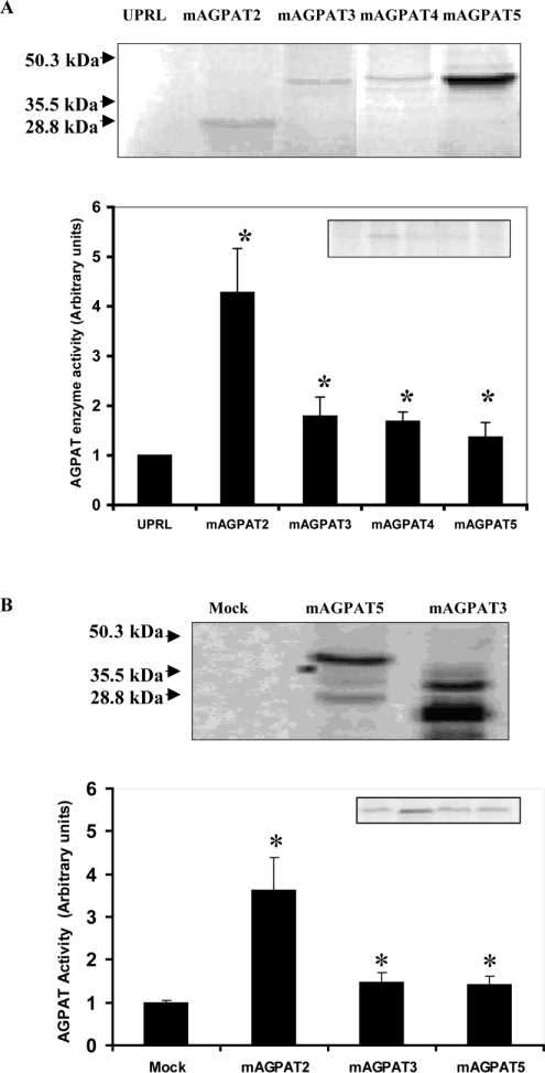
Figure 2. Expression of mAGPAT in vitro and in vivo and enzyme activities of mAGPATs.
(A) Expression of mAGPAT2, mAGPAT3, mAGPAT4 and mAGPAT5 protein using the in vitro protein translation system. Reticulocyte lysates were prepared and proteins were [35S]methionine radiolabelled and visualized by exposure to X-ray film (upper panel). UPRL, unprocessed lysate. AGPAT enzyme activities were determined as described in the Materials and methods section (lower panel). (B) Expression of V5-His epitope tag fused to the C-terminus of mAGPAT3 and mAGPAT5 in COS-1 cells (upper panel). Appearance of the mAGPAT5 band is greater since the protein contains 13 methionine residues instead of 11 and 7 methionine residues in mAGPAT2 and mAGPAT3 respectively. Molecular mass markers are indicated on the left. An equal amount of protein from cell lysate was used to determine relative mAGPAT enzyme activitities as described in the Materials and methods section (lower panel).

