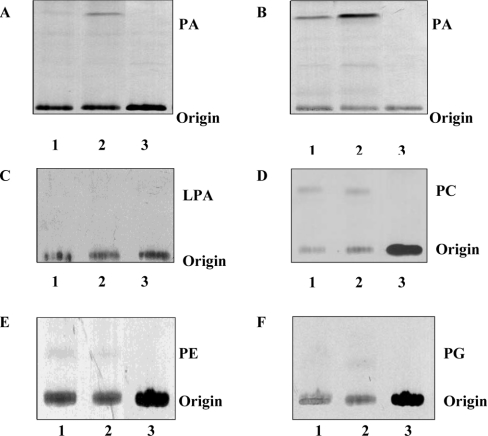
Figure 3. Specificity of mAGPAT2 activity.
AGPAT activities of mAGPAT2 from in vitro (A) and in vivo (B) sources. The film exposure time was 24 h for (A) and (B). (A) Lane 1, unprogrammed reticulocyte lysate; lane 2, reticulocyte lysate containing mAGPAT2; lane 3, [1-14C]oleoyl-CoA. (B) Lane 1, COS-1 cell lysate; lane 2, COS-1 cell lysate from mAGPAT2 transfected cells; lane 3, [1-14C]oleoyl-CoA. (C) GPAT activities in reticulocyte lysates. (D) LPCAT activities in reticulocyte lysates. (E) LPEAT activities in reticulocyte lysates. (F) LPGAT activities in reticulocyte lysates. Lane 1, unprogrammed reticulocyte lysate. Lane 2, reticulocyte lysate containing mAGPAT2. Lane 3, radiolabelled substrate [1-14C]oleoyl-CoA. The film was exposed for 72 h. Representative autoradiographs are depicted.

