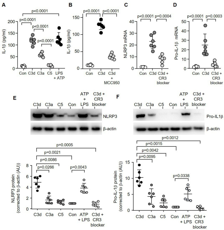
Figure 2.

C3d signalling via CR3 triggers NLRP3 inflammasome activation and IL-1β release. Monocytes remained untreated (Con) or challenged with C3d, C3a, C5 (10 µg/mL) or ATP (3 mM) and LPS (50 ng/mL) (A), or MCC950 (1 µM) (B), and extracellular levels of IL-1β determined by ELISA (n=6 biological repeats, one-way ANOVA, followed by Tukey’s posthoc multiple comparison test). (C&D) NLRP3 (C) or pro-IL-1β (D) Real-Time quantitative Polymerase Chain Reaction of monocyte mRNA (fold change) post C3d treatment (10 µg/mL) with or without CR3 blocking antibody (C3d blocker, n=6 biological repeats, one-way ANOVA, followed by Tukey’s posthoc multiple comparison test). HC monocytes (1×10⁵) were incubated with C3d, C3a, C5 or ATP and LPS. Cell lysates were collected and western blotted for NLRP3 (E) or pro-IL-1β (F). All results are expressed as relative densitometry units (DU), with representative western blots presented (n=6 biological repeats, one-way ANOVA, followed by Tukey’s posthoc multiple comparison test). HC, healthy controlIL-1β, interleukin-1β; LPS, lipopolysaccharide; NLRP3, NOD-, LRR- and pyrin domain-containing protein 3.
