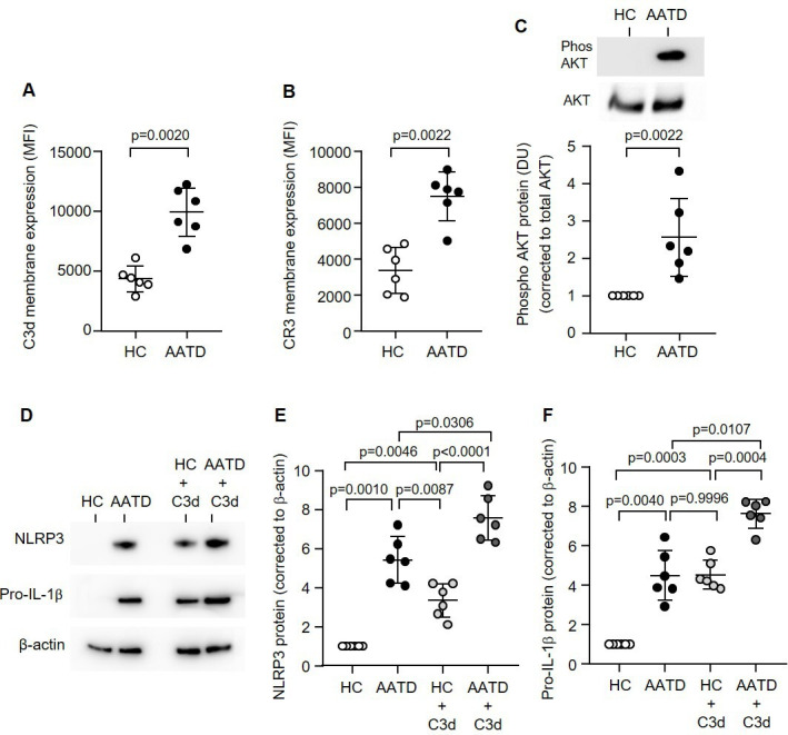
Figure 4.

The CR3:C3d signalling pathway is increased in the monocytes of patients with AATD. C3d (A) or CR3 (B) levels were measured on the outer plasma membranes of monocytes isolated from AATD or HC by flow cytometry (n=6 subjects per group, non-parametric Mann-Whitney U test). (C) Expression levels of AKT in AATD or HC subjects measured by western blot analysis of monocyte whole cell lysates. Phosphorylation levels were normalised to respective total protein. Results are expressed as DU, with representative western blots presented (n=6 subjects per group, non-parametric Mann-Whitney U test). (D–F) NLRP3 (D and E) or pro-IL-1β (D and F) protein was detected by western blot analysis in HC or AATD monocytes either untreated or treated with C3d (10 µg/mL). Data are expressed as relative DU, with representative western blots presented (n=6 biological repeats, one-way ANOVA, followed by Tukey’s posthoc multiple comparison test). AATD, alpha-1 antitrypsin deficiency; DU, densitometry units; HC, healthy controls; NLRP3, NOD-, LRR- and pyrin domain-containing protein 3.
