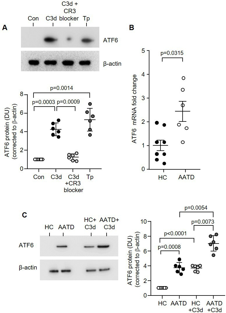
Figure 5.

CR3 inhibition modulates C3d-induced ER stress in AATD monocytes. (A) Western blot and densitometry analysis of cleaved ATF6 expression in control (Con) untreated monocytes from HC or following treatment with C3d (10 µg/mL) for 16 hours. Negative and positive controls included preincubation with CR3 blocker (clone mAb 107, 1 µg/mL) and treatment with the ER stress inducer thapsigargin (Tp) (100 nM), respectively (n=6 biological repeats per group, one-way ANOVA). (B) ATF6 qRT-PCR of HC (n=8) or AATD (n=6) monocyte mRNA revealed significantly increased expression in AATD (non-parametric Mann-Whitney U test). (C) ATF6 expression in resting HC monocytes compared with AATD cells, and following C3d challenge (n=6 subjects per group, one-way ANOVA, followed by Tukey’s posthoc multiple comparison test). AATD, alpha-1 antitrypsin deficiency; ER, endoplasmic reticulum; HC, healthy individuals.
