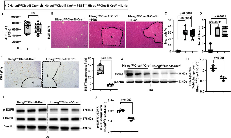
Figure 6.
Liver repair after IR injury is impaired when hb-egf is deleted in hepatic macrophages. Male Hb-egffl/flClec4f-Cre+/-mice and WT littermates (Male Hb-egffl/flClec4f-Cre-/-) were subjected to hepatic IR surgery. After 24h, Hb-egffl/flClec4f-Cre+/- mice were divided into two groups and injected (i.p.) with PBS or IL-4c (n=6/group). (A) Serum ALT levels at 6h after IR surgery. (B, C) Liver necrosis (N, outlined areas) was evaluated and quantified on day 7 after IR surgery. (D) Liver pathology was assessed by using the Suzuki’s scoring system on day 7 after IR surgery. (E, F) Proliferative hepatocytes were stained for Ki67 and quantified on day 3 after IR surgery (n=4/group). (G, H) PCNA protein expression was detected by Western blotting and quantified. (I, J) p-EGFR and t-EGFR protein expression levels were detected by Western blotting and quantified. Two-tailed unpaired Student’s t-test with Welch’s correction was performed in A, F, H and J. One-way ANOVA was performed in C and D.

