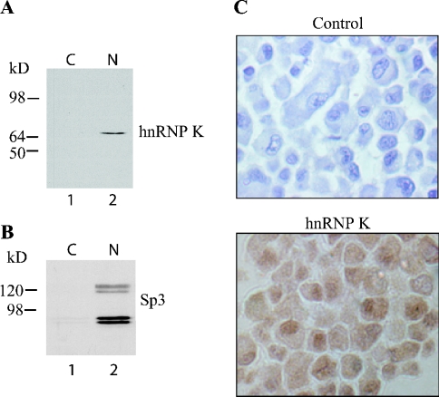
Figure 4. HnRNP K is expressed in osteoblastic cells.
(A) Cytosolic and nuclear extracts from ROS 17/2.8 cells were immunoblotted and probed with an anti-hnRNP K antibody, revealing expression primarily in the nucleus. (B) Immunoblotting with Sp3 reveals the specificity of the nuclear fraction. (C) Immunocytochemistry of ROS 17/2.8. Note that ROS17/2.8 cells show immunoreactivity (brown stain) for hnRNP K, which is more abundant in the nucleus than in the cytosol. In control cells, the primary antibody was omitted (×200 magnification).

