
FIGURE 4.
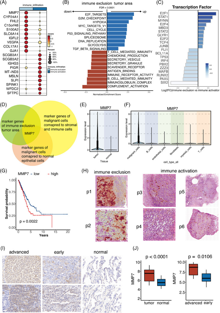
MMP7 was highly expressed in tumour area with infiltrated APOE+ tumour‐associated macrophages (CD14+ cells). (A) The differentially expressed genes (DEGs) of tumour cells in samples with distinct immune infiltration patterns. (B) Pathway analysis unveiled distinct biological activities in immune activation and immune exclusion tumour areas. (C) Expression patterns of the most varied transcription factors (TFs) in two types of tumour areas. (D) MMP7 was identified as the potential tumour marker correlating with the infiltrated CD14+APOE+ cells. (E) MMP7 exhibited elevated expression in tumour cells compared to normal epithelial cells. (F) MMP7 exhibited elevated expression in tumour cells compared to immune cells and stromal cells. (G) Patients with high MMP7 levels had shorter survival time. (H) The spatial expression patterns of MMP7 in two immune infiltration types of samples. (I) Immunohistochemistry images of MMP7 in our cohort consisting of 60 non‐small cell lung cancer (NSCLC) patients. (J) Boxplots displaying the distribution of MMP7 in normal, early and advanced stage samples.