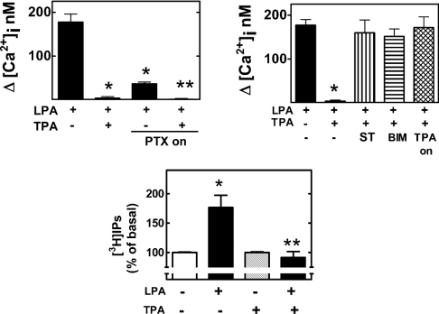
Figure 2. Effects of PKC and the roles of pertussis-toxin-sensitive G-proteins in the actions of LPA.
Cells were incubated with the agents indicated as described in the Materials and methods section. LPA, 1 μM LPA; TPA, 1 μM PMA; TPA-on, PMA (1 μM, incubated overnight); PTX-on, pertussis toxin 100 ng/ml overnight; ST, preincubation with 300 nM staurosporine before the addition of other agents; BIM, preincubation with 1 μM bisindolylmaleimide I before the addition of other agents. Mean values are plotted and vertical lines represent the S.E.M. for 6–12 experiments using different cell preparations. Upper left panel: *P<0.001 versus LPA alone, **P<0.001 versus LPA alone and P<0.05 versus LPA treated with pertussis toxin. Upper right panel: *P<0.001 versus all the other treatments. Lower panel: *P<0.01 versus basal (no treatment); **P<0.01 versus LPA without PMA.

