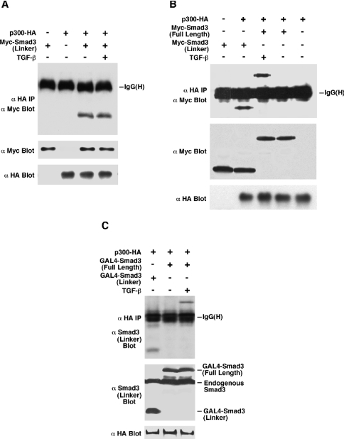
Figure 3. The linker region of Smad3 physically interacts with p300.
(A) and (B) HEK-293 cells were co-transfected with Myc-Smad3 (Linker), Myc-Smad3 (Full Length), p300–HA and constitutively active TGF-β type I receptor TβRI (T204D) for TGF-β induction. Cell lysates were immunoprecipitated (IP) by HA antibody, followed by immunoblotting with an antibody against the Myc epitope. Lysate controls are shown for Myc-Smad3 (Linker) and Myc-Smad3 (Full Length) expression levels by Myc immunoblotting, and p300–HA levels by HA immunoblotting. (C) HEK-293 cells were co-transfected with GAL4–Smad3 (Linker), GAL4–Smad3 (Full Length), p300–HA and TβRI (T204D) for TGF-β induction. Cell lysates were immunoprecipitated by HA antibody, followed by immunoblotting with an antibody against the Smad3 linker region. GAL4–Smad3 (Linker) and GAL4–Smad3 (Full Length) expression levels were examined by the antibody against Smad3 linker region. p300–HA levels were examined by HA immunoblotting.

