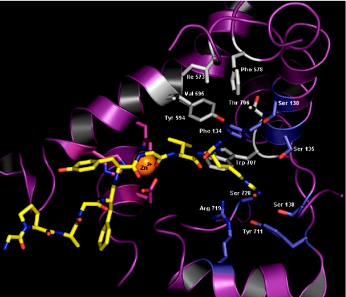
Figure 8. Model of D. melanogaster NEP2 with LomTK-1 docked at the active site.
A ribbon diagram showing the active-site space with substrate and metal ion co-ordinating residues. The peptide substrate LomTK-1 is shown in yellow and the ligand-binding subsites S1′ and S2′ are shown with their side-chains in grey and light blue respectively. Associated helices and zinc co-ordinating ligands are shown in magenta.

