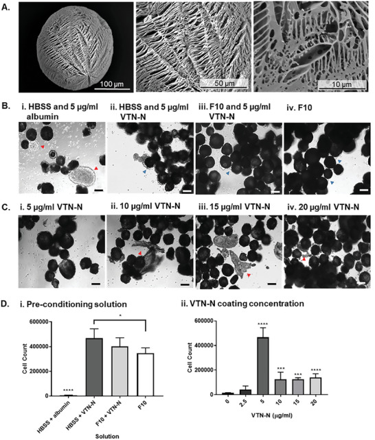
Figure 1.

Optimization of TIPS microcarriers for iPSC attachment. A) Ultrastructural features of the 2% 7507 TIPS microcarriers were assessed using SEM. B) and C) Conditions for pre‐conditioning TIPS microcarrier to facilitate iPSC attachment. 5 × 10⁵ P6 iPSC were seeded on 20 mg of microcarriers pre‐conditioned with either HBSS and 5 µg/ml albumin (negative control), HBSS and 0–20 µg/ml VTN‐N, complete F10 media and 5 µg/ml VTN‐N and complete F10 media (positive control). iPSC were attached under static dynamic conditions (30 seconds gentle agitation at 30 RPM every hour) for 24 hours. D) After 24 hours, iPSC attachment to the TIPS microcarriers was quantified using a NucleoCounter NC‐200 automated cell counter. Red arrows show cellular clumping and uneven attachment of cells to the microcarriers. Blue arrows show cell attachment to the microcarriers, with formation of cellular connections between microcarriers. Scale bars represent 100 µm. Data are presented as mean ±SD. The significance of the data was calculated by two‐way ANOVA, Tukey's post‐hoc correction. (n = 6, *P ≤ 0.05, ***P ≤ 0.001, ****P ≤ 0.0001).
