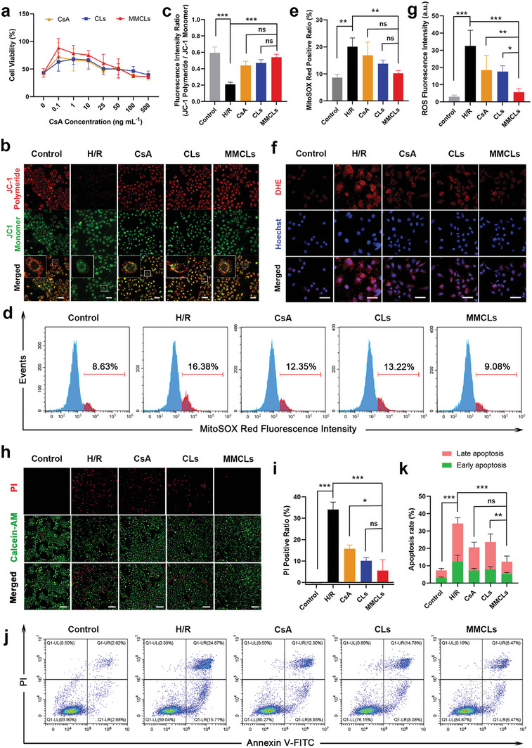
Figure 4.

Verification of the protection effect of MMCLs on H/R‐injured AML12 cells in vitro. a) Cell viability of H/R‐injured cells after being treated with free CsA, CLs, and MMCLs at CsA concentrations of 0 − 500 ng mL−1 as evaluated by CCK‐8 assay. b) JC‐1 mitochondrial membrane potential in normal cells and H/R‐injured cells following the treatment with CsA, CLs, and MMCLs as imaged by CLSM (scale bar: 50 µm), and c) the corresponding fluorescence intensity ratio of JC‐1 polymeride and monomer (n = 5). d) Flow cytometry analysis of the superoxide levels using MitoSOX Red staining, and e) the corresponding positive ratio of MitoSOX Red (n = 5). f) CLSM images of AML12 cells stained by DHE (scale bar: 50 µm), and g) the corresponding fluorescence intensity of intracellular ROS (n = 5). h) HCI analysis of cell death using PI staining (scale bar: 200 µm) and i) the corresponding positive ratio of PI (n = 5). j) Flow cytometry analysis of early and late apoptosis by annexin V‐FITC/PI dual staining assay, and k) the corresponding rates of early and late apoptosis (n = 5). Data are presented as mean ± SD. The statistical significance was analyzed using one‐way ANOVA following Tukey's multiple comparisons test (*p < 0.05; **p < 0.01; ***p < 0.001, ns = no significance).
