Figure 3.

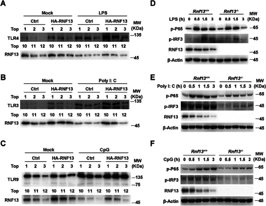
RNF13 enhances endosomal TLRs signaling activity. A) Western blot assay of Top1‐3 and Top10‐12 layers in subcellular fraction assay with control (Ctrl) and RNF13 overexpressing (HA‐RNF13) RAW264.7 cells treated with LPS (100 ng mL−1) for 0 or 3 h. B) Western blot assay of Top1‐3 and Top10‐12 layers in subcellular fraction assay with control (Ctrl) and RNF13 overexpressing (HA‐RNF13) RAW264.7 cells treated with Poly I: C (10 µg mL−1) for 0 or 3 h. C) Western blot assay of Top1‐3 and Top10‐12 layers in subcellular fraction assay with control (Ctrl) and RNF13 overexpressing (HA‐RNF13) RAW264.7 cells treated with CpG (2 µM) for 0 or 3 h. D) Western blot assay of TLR4 signaling pathway in Rnf13 +/+ and Rnf13 −/− RAW264.7 cells treated with LPS (100 ng mL−1) for indicated hours. E) Western blot assay of TLR3 signaling pathway in Rnf13 +/+ and Rnf13 −/− RAW264.7 cells treated with Poly I: C (10 µg mL−1) for indicated hours. F) Western blot assay of TLR9 signaling pathway in Rnf13 +/+ and Rnf13 −/− RAW264.7 cells treated with CpG (2 µM) for indicated hours. Data are representative of three independent experiments (A–F).
