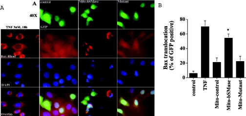
Figure 3. Overexpression of the mitochondria-targeted bSMase-D295G mutant did not cause Bax translocation.
MCF7 cells were treated with 3 nM TNFα for 18 h or were co-transfected with 3.5 μg of pCMV/GFP/Mito empty vector plus 0.35 μg of pEGFP-N1 vector, or with 3.5 μg of pCMV/bSMase-GFP/Mito vector plus 0.35 μg of pEGFP-N1 vector, or 3.5 μg of pCMV/bSMase-D295G-GFP/Mito mutant vector plus 0.35 μg of pEGFP-N1. At 48 h after transfection, cells were fixed, immunostained with anti-Bax mouse monoclonal antibody, and visualized with rhodamine-conjugated secondary antibody. Analysis and quantification of Bax translocation was performed by indirect immunofluorescence as described in the Experimental section. Data are from one experiment performed in duplicate, representative of at least three separate experiments. Asterisks indicate a significant difference (P<0.05) compared with the control. For statistical analysis, Student's t test for paired sample means was used.

