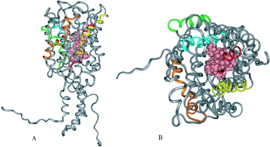
Figure 7. GLUT4 3D structure.
(A) Ribbon representation (side view) of GLUT4 3D structure, showing the QLS site (pink) in CPK arrangement. C- and N-terminal regions are shown as a coil due to lack of a suitable template. (B) Extracellular top view in ribbon representation of GLUT4 3D structure. The QLS site, at the pore region in CPK arrangement (pink), comprises residues involved in glucose transport. Transmembrane helices are coloured: helix 2 (orange), helix 5 (yellow), helix 7 (red), helix 10 (green) and helix 11 (cyan).

