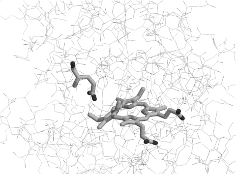
Figure 3. Three-dimensional structure of B. subtilis ferrochelatase co-crystallized with N-methylmesoporphyrin.
The conserved Glu-264 (Glu-289 in murine ferrochelatase) and the bound porphyrin are highlighted. The image of the porphyrin–ferrochelatase complex (accession code 1C1H from the Protein DataBank) was generated using RasMol 2.7.2.1.

