Figure 1.
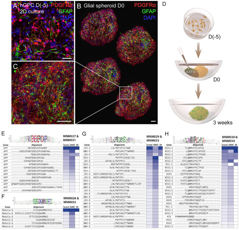
Workflow for glial spheroid formation and transduction with BRAVE library. (A) 2D culture of hGPCs expressing canonical glial markers (GFAP, PDGFRα) by immunocytochemistry. (B,C) Confocal immunofluorescence images of (B) four cryosections from a glial spheroid expressing glial markers on the day of AAV transduction (B), with magnification (C). (D) Schematic illustrating glial spheroid formation and viral transduction with corresponding time points. (E–H) Shared sequence homology and consensus motifs for the MNM027-MNM031 (E), MNM028-MNM032 (F), MNM029-MNM033 (G), and MNM030-MNM034 (H) variants. Data information: 2D, two-dimensional; hGPCs, hESC-derived glial progenitor cells; D, day. Scale bars 50 μm.

