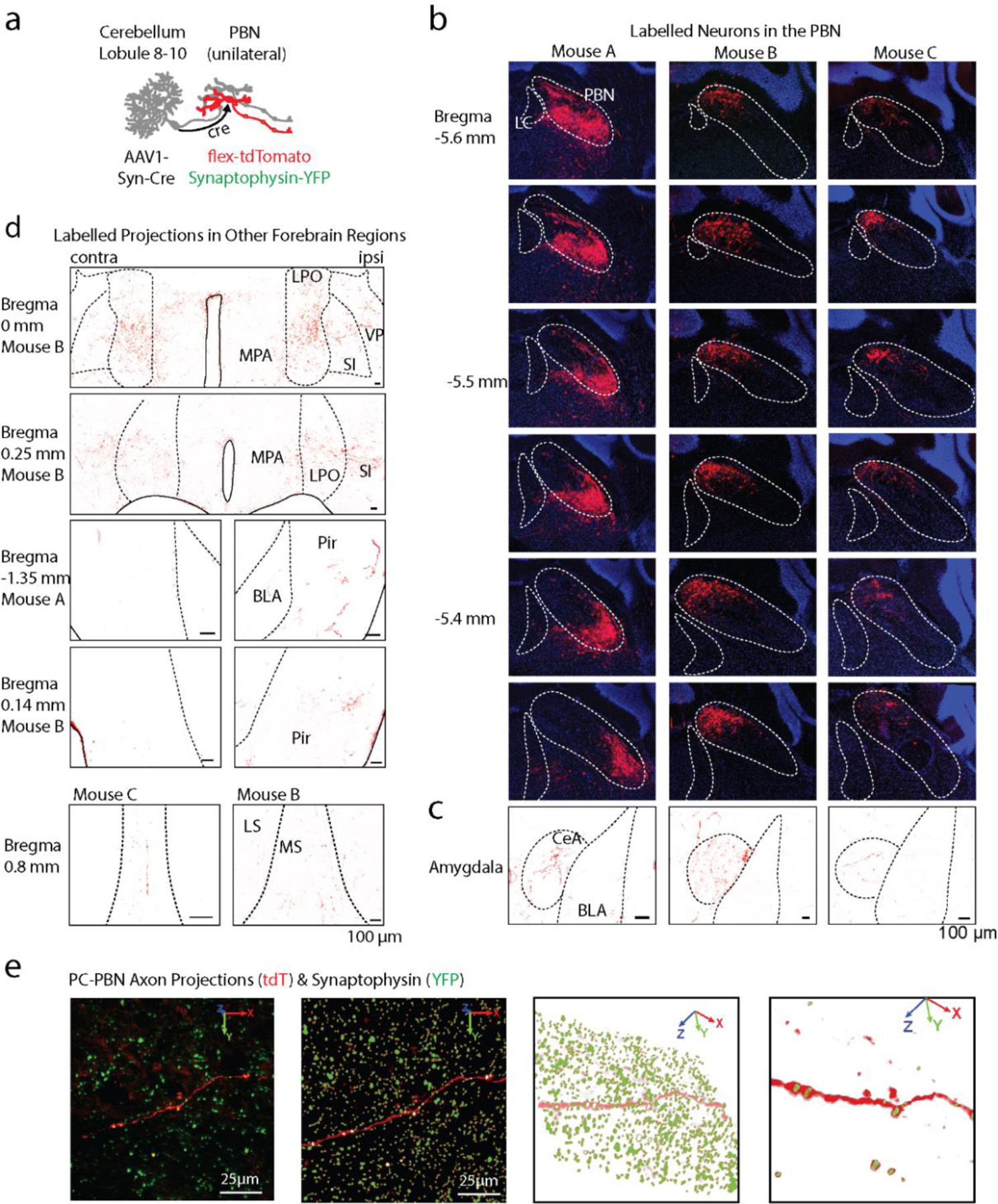
Extended Data Fig. 7. Labelling of the PC-PBN projection pathway.

A. An anterograde AAV-cre was injected into the posterior cerebellar vermis, and AAVs with cre-dependent tdTomato were injected into the PBN. This led to tdT-expression in PC-recipient PBN neurons. The PBN of mouse C was also injected with AAV1-Synaptophysin-YFP to label the presynaptic boutons of all PBN neurons.
B. A series of sections shows that tdT expression was restricted to the PBN of the three mice injected as in a. Approximate location of the LC and PBN is indicated, as determined by the mouse brain atlas and DAPI staining.
C. tdT-expressing axons are shown in the amygdala of each mouse.
C. tdT-expressing axons are shown in the indicated regions for the indicated mice.
D. To visualize PC-PBN projections and determine synaptophysin overlap, 20 μm confocal stacks of each region were taken (left panel), and synaptophysin and axon signals were segmented out (right panels). Puncta associated with a tdTomato-expressing axon are displayed in the examples for the indicated regions.
