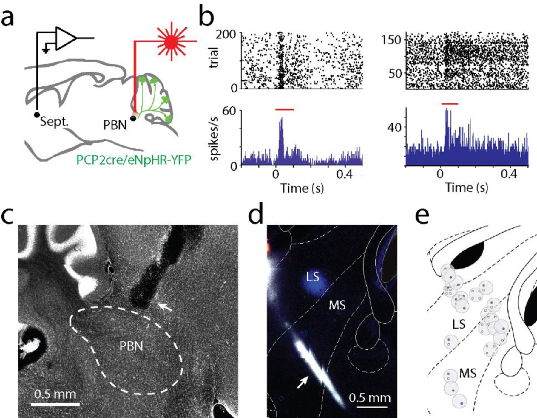
Extended Data Fig. 8: Suppressing the PC-PBN pathway rapidly increases firing in the septum.

a. In awake, head-restrained mice expressing halorhodopsin in PCs, the PC-PBN pathway was optically suppressed and single-unit recordings were made in the septum.
b. Two example cells showing increases in firing during the 100 ms light pulse (red line).
c. Optical fiber implant into the parabrachial nuclei. Shown is a sagittal slice of the PBN with cerebellum on the left. Optical fiber implant is indicated with the white arrow. Boundaries of the PBN are indicated with white dotted lines.
d. Example recording site through the medial septum (MS) and lateral septum (LS). Probes were coated with different color dyes to enable post-hoc verification of recording sites. Recording sites were determined by matching the dye with the final position of the probe inside the brain. Dye is shown in white.
e. Recording sites for all experiments shown above. Every grey circle indicates a recording site. Blue dots indicate responding cells and black dots indicate nonresponding cells for each site.
