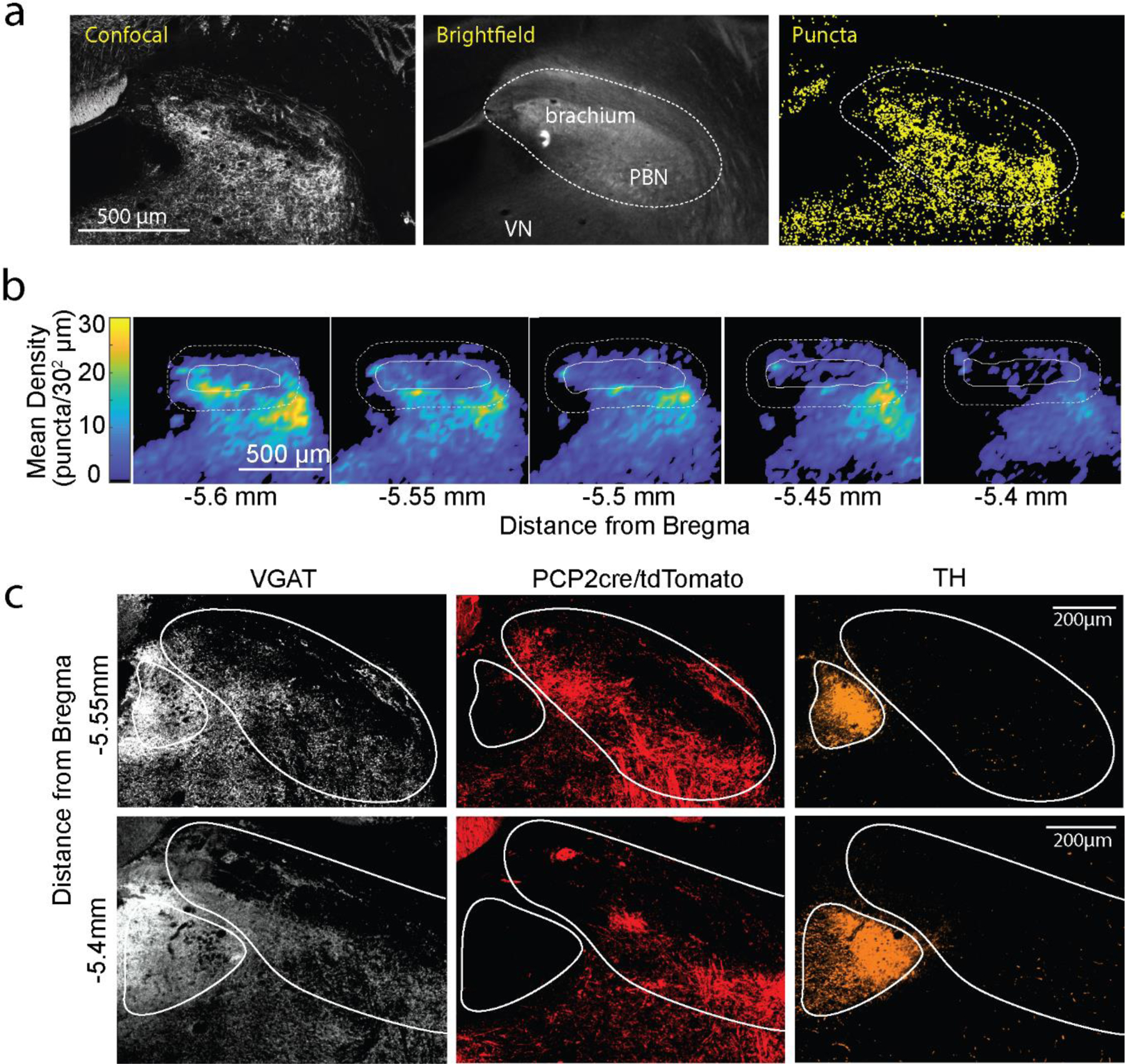
Extended Data Fig. 2: Purkinje cell synapses within the PBN.

a. Method for registering PC synapses and identifying the PBN in the PCP2cre/Synaptophysin-tdTomato mouse. The confocal image (left) showing tdTomato fluorescence, the brightfield image (middle) with the brachium conjunctivum noted, and identified presynaptic boutons (right, yellow) are shown with the bounds of the PBN delineated.
b. Average heatmaps of identified PC Synaptophysin-tdT puncta in the PBN. Individual slices were binned at 30 μm2 and averaged by aligning the center of the brachium. Each heatmap is an average of 4 slices (from 2 animals, 2 hemispheres per animal). The bold white line outlines the brachium, and the dotted line the PBN.
c. Immunohistochemistry was used to identify inhibitory synapses (vGAT, white), and the locus coeruleus (tyrosine hydroxylase, orange) in a PCP2cre/tdTomato mouse where PC axons and boutons were labelled (tdT, red). Very few PC boutons were apparent in the locus coeruleus.
