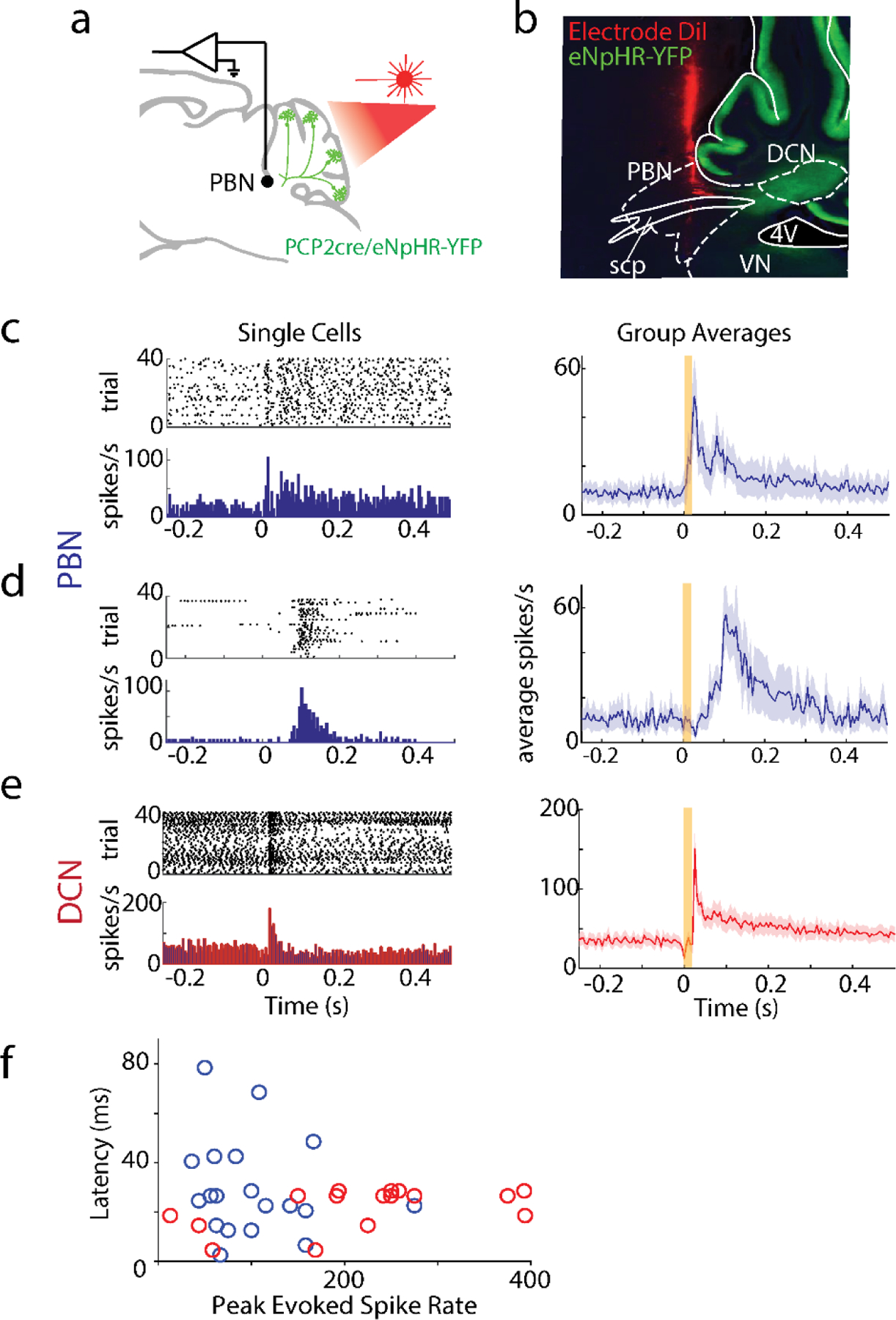
Extended Data Fig. 3: PBN neurons rapidly increase firing in response to suppression of PC firing.

A. Single-unit, multielectrode array recordings were made in the PBN or DCN in awake, head-restrained PCP2cre/Halo mice (n=6). The posterior cerebellar cortex was stimulated through a thinned skull (20 ms, red light).
B. Recording sites were recovered by coating the silicon probe with DiI. An example electrode tract is shown. scp: superior cerebellar peduncle, 4V: 4th ventricle
C. left, Firing evoked in a rapidly responding PBN neuron (<30 ms latency). right, Summary of rapidly responding PBN neurons (13/28 neurons). Shaded area indicates standard error.
D. Same as C but for slower responding PBN neurons (6/28 neurons).
F. Same as C but for DCN neurons (16/16 neurons).
F. Latencies of PBN neurons (blue) and DCN neurons (red) as a function of evoked firing rate.
