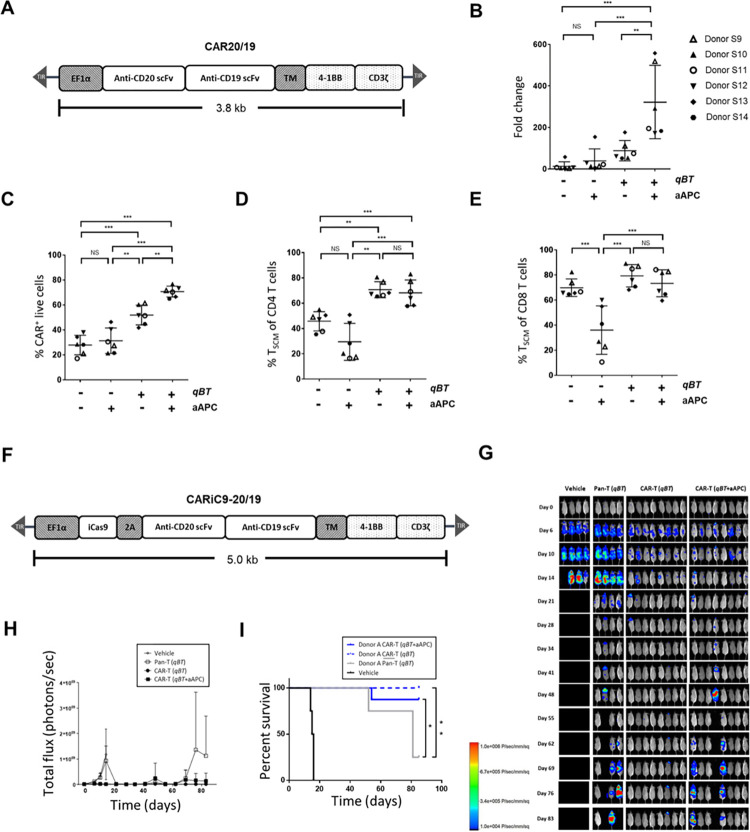
Fig 1. Effect of qBT and aAPC on CAR-T cells.
(A) Schematic diagram of the CAR20/19 CAR construct. (B-E) Human peripheral blood mononuclear cells (PBMC) electroporated with Quantum pBac™-expressing CAR and cultured for 10 days in the presence or absence of aAPC and/or Quantum Booster™ (qBT) were harvested and assessed for (B) cell expansion fold change, (C) percentage CAR+ of live cells (CD3+ PI-, >90%), and percentage of TSCM cell subsets in (D) CD4+ and (E) CD8+ cells. Data shown are from six healthy donors. Horizontal lines represent the mean and s.e.m. fold change (B), mean and s.e.m. percentage of CAR+ cells (C), and mean and s.e.m. percentage of TSCM cell subsets (D and E). * p < 0.05, ** p < 0.01, *** p < 0.001. (F) Schematic diagram of the CARiC9-20/19 CAR construct. A T2A sequence enables co-expression of iCasp9 and CD20/CD19-targeted scFvs under the EF1α promoter. (G-I) In vivo functional characterization of CAR-T cells produced by perfusion culture system. (G) In vivo cytotoxicity of CAR-T cells with or without pre-incubation with aAPC in Raji-GFP/Luc-bearing immunodeficient mice. Fluorescence intensity values (H) and survival curves (I) of mice from (G) plotted against time. Results shown are from 4 to 8 mice/group. T cells were obtained from a representative donor. Vehicle and Pan-T cells (non-engineered T cells) were used as controls. qBT was present in all cell culture conditions. Groups were compared by log-rank (Mantel-Cox) test, *p < 0.05, **p < 0.01.

