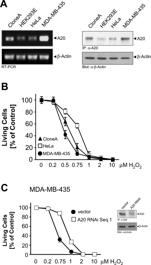
Figure 1. Cells which express high basal levels of A20 are less protected against H2O2-induced death.
(A) RNA was isolated from the indicated cell lines and RT-PCR was performed for A20 and β-actin, or A20 was immunoprecipitated and revealed by immunoblotting with anti-A20 (α-A20). Immunoblots for β-actin (α-β-actin) served as a control. (B) HeLa cells (□), Clone A cells (▲) and MDA-MB-435 cells (●) were treated with the indicated doses of H2O2 for 16 h. Living cells were counted by Crystal Violet staining. Results are means±S.D. (C) MDA-MB-435 cells transfected with control pSUPER (□) or transfected with pSUPER.A20 RNAi for 48 h (●) were treated with the indicated doses of H2O2 for 16 h. Cell viability was determined by Crystal Violet staining. Results are means±S.D. Inset, A20 was immunoprecipitated and revealed by immunoblotting with anti-A20 (α-A20). Immunoblots for β-actin (α-β-actin) served as a control.

