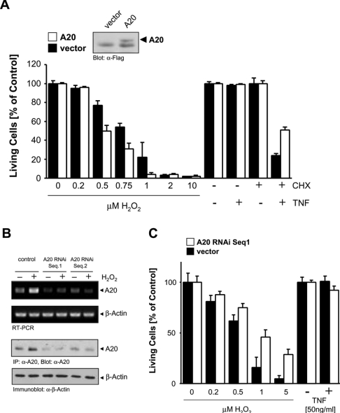
Figure 2. A20 sensitizes cells to oxidative-stress-mediated cell death.
(A) HeLa cells were transfected with A20 (open bars) or vector control (closed bars). At 48 h after transfection, cells were treated with H2O2 or TNF-α (50 ng/ml)/CHX (1 μg/ml) as indicated for 16 h. Living cells were analysed by Crystal Violet staining. Results are means±S.D. A20 expression was shown by immunoblotting (α-FLAG, the lower band is non-specific). (B) HeLa cells were transfected with vector control (pSuper) or A20 RNAi (pSuper.A20 RNAi Seq.1 or Seq.2) for 48 h, and were then treated with H2O2 (500 nM, 4 h). A20 was immunoprecipitated and immunoblotted (α-A20) or RNA was isolated and RT-PCR was performed. β-Actin expression served as a control (immunoblot: α-β-actin, or RT-PCR). (C) HeLa cells were transfected with vector control (pSuper, closed bars) or A20 RNAi (pSuper.A20 RNAi Seq.1, open bars) for 48 h, and were then treated with increasing concentrations of H2O2 or with TNF-α, as indicated. Living cells were analysed by Crystal Violet staining. Results are means±S.D.

