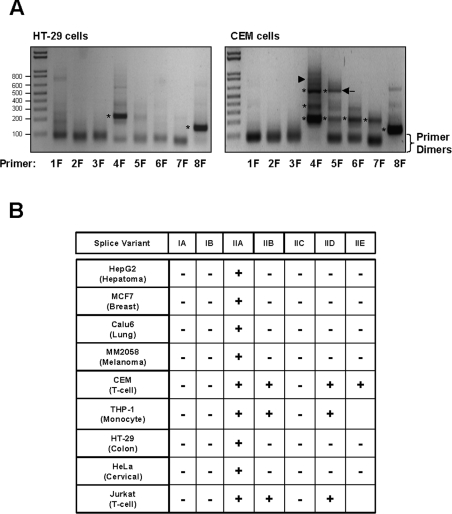
Figure 2. Identification of RNA splice variants in human cell lines.
Forward primers (1F–8F specific for exons 1–8 respectively) were designed to amplify the different exons using a common reverse primer (ExonR) in the coding region. Amplification gave a series of products depending on the combination of exons. Predicted product sizes for the different RNA variants are listed in Table 3. (A) Representative electrophoretic patterns from cells that express only one variant, Type IIA (left-hand panel). A band can be seen with the 4F and 8F primers, indicating the presence of exons 4 and 8. The right-hand panel shows the electrophoretic pattern for cells expressing several variants. In this case (CEM cells), multiple bands were seen with primers 4F and 5F, while a single band was seen with 6F, 7F and 8F. From the sizes, the different RNA variants can be deduced. Bands indicated with the asterisks were cloned and sequenced to confirm their identity. In lane 5F, an unknown band of approx. 550 bp was present (arrow). When sequenced, this band represented a new variant containing exons 5, 6, 8 and 9. (B) Occurrence of the different RNA species in human cell lines. Variant IIA was present in all cells, while only the blood-derived lines showed evidence of multiple RNA species.

