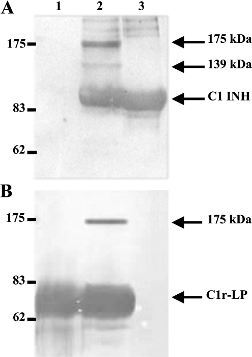
Figure 8. Western blots of C1r-LP/C1 INH mixtures.
Purified recombinant C1r-LP was incubated with an equimolar amount of C1 INH at 37 °C for 1 h and duplicate aliquots of the mixture were subjected to SDS/PAGE (7.5% gel) under reducing conditions, followed by Western blotting. The upper blot (A) was developed with anti-C1 INH and the lower blot (B) with anti-C1r-LP serum. Both blots were developed with peroxidase-conjugated anti-rabbit IgG, followed by chemiluminescent reagents (A) or 4-chloro-1-naphthol (B). Lane 1, purified C1r-LP; lane 2, C1r-LP/C1 INH mixture; lane 3, C1 INH. Positions of molecular mass markers are on the left. Arrows indicate the uncomplexed proteins and the two observed complexes of 175 and 139 kDa.

