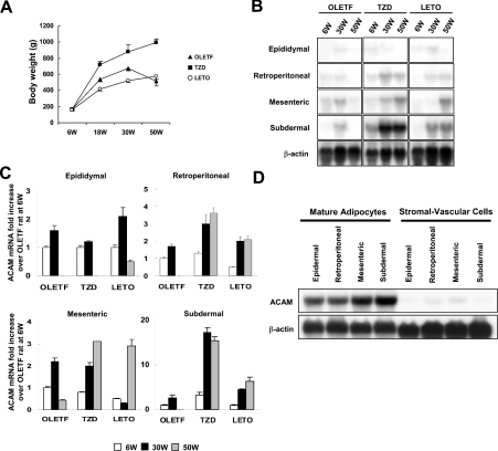
Figure 4. Northern blot analyses of ACAM in WATs of OLETF rats.
(A) The body mass of OLETF rats, TZD rats [OLETF rats with free access to chow containing pioglitazone (approx. 1 mg/kg of body mass per day] and LETO rats are shown. (B) Total RNAs (20 μg) isolated from various WATs of 6-week (6W), 30-week (30W) and 50-week (50W)-old rats were subjected to Northern blot analyses. (C) For each adipose tissue, the fold increase in the ACAM mRNA/β-actin ratio compared with OLETF rats at 6 weeks of age is indicated. Each value is expressed as the mean±S.D. (D) Mature adipocytes and stromal vascular cells were isolated by collagenase digestion and ACAM mRNA expression was analysed with Northern blot analysis. ACAM mRNA is predominantly expressed in mature adipocyte fractions and barely detected in stromal vascular cells.

