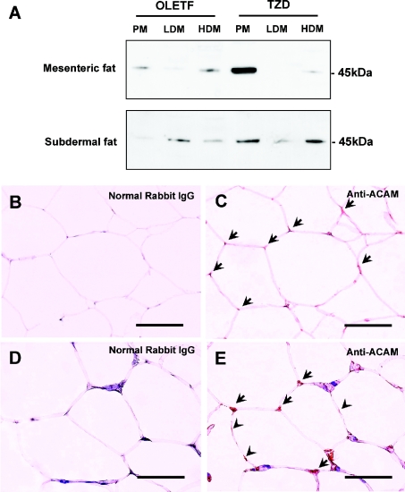
Figure 5. Western blot and immunohistochemistry of ACAM in WATs of OLETF rats.
(A) Mesenteric and subdermal WATs derived from OLETF and TZD rats at 30 weeks of age were subjected to subcellular fractionation of adipocytes: plasma membrane (PM), HDM and LDM fractions. Anti-ACAM antibody reveals approx. 45 kDa major bands in plasma membrane fraction of mesenteric and subdermal adipose tissues of OLETF rats. Pioglitazone up-regulates the ACAM protein expression in mesenteric and subdermal adipose tissues of OLETF rats. In HDM and LDM fractions, faint bands at approx. 45 kDa were detected. (B–E) Immunostaining of ACAM in subdermal adipose tissues of 30-week-old OLETF rats (C) and TZD rats (E). Normal rabbit IgG was incubated with subdermal adipose tissues of OLETF rats (B) and TZD rats (D) as a negative control. In subdermal adipose tissues in TZD rats, faint immunoreactivity of ACAM is detected on the surface of adipocytes [arrowheads in (E)] and somehow more intense staining is observed at corners of polygonal-shaped adipocytes [arrows in (E)]. Scale bars, 100 μm.

