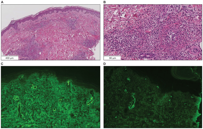
Figure 3.
Skin biopsy findings on the lower leg. (A, B) Hematoxylin and eosin staining showed neutrophil-dominated inflammatory cell infiltration from the small vessel walls into the surrounding tissues (scale bar [A] = 400 µm, [B] = 90 µm). (C) Immunofluorescence staining revealed granular IgA deposition in the vessel walls (×200). (D) Immunofluorescence staining revealed granular C3 deposition in the vessel walls (×200).

