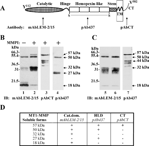
Figure 1. Characterization of soluble MT1-MMP forms.
(A) Domain structure of active MT1-MMP with the domain specific antibodies used in the present study. (B) Media from BS-C-1 cells expressing MT1-MMP in the absence (lane 1) or in the presence of 2 μM TAPI-1 (lanes 2–4) were concentrated and subjected to SDS/PAGE (15% reducing gel) followed by immunoblot analysis using the mAbLEM-2/15 to the catalytic domain (lanes 1 and 2), pAbCT to cytoplasmic tail (lane 3) and pAb437 to the haemopexinlike domain (lane 4) of MT1-MMP. (C) Media of MT1-MMP expressing BS-C-1 cells obtained in the presence of 2 μM TAPI-1 were collected (lane 5) and subjected to ultracentrifugation (supernatant, lane 6; pellet, lane 7). The samples were subjected to SDS/PAGE (15% reducing gel) followed by immunoblot analysis using the mAbLEM-2/15 to the catalytic domain (lanes 5 and 6) and pAb437 to the haemopexin-like domain (lane 7). (D) Summary of the species of soluble forms of MT1-MMP detected in the media of BS-C-1 cells expressing MT1-MMP and their recognition by various anti-MT1-MMP antibodies. Cat.dom., catalytic domain; CT, C-terminus; HLD, haemopexin-like domain; TM, transmembrane.

