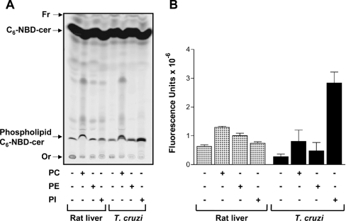
Figure 2. Fluorescent phosphosphingolipids synthesized by rat liver and epimastigote microsomal membranes.
(A) Membranes (100 μg·ml−1) were incubated in 100 μl of 100 mM Tris/HCl (pH 7.4) containing 0.1 mM C6-NBD-cer, 6 mg·ml−1 fatty-acid-free BSA (rat liver) or 0.1% TX-100 (T. cruzi), in the absence (−) or presence (+) of 1 mM PC, PE or PI. After 30 min of incubation at 37 °C (rat liver) or 28 °C (T. cruzi), lipids were extracted, separated by TLC and visualized using a PhosphorImager Storm 860 (Molecular Dynamics). The relative positions of synthesized phosphosphingolipids labelled with C6-NBD-cer and unchanged C6-NBD-cer are indicated on the left, together with the origin (Or) and the front (Fr) of the chromatogram. (B) Fluorescence intensities of synthesized phosphosphingolipids containing C6-NBD-cer from three independent experiments (means±S.E.M.) were quantified using ImageQuant5.2.

