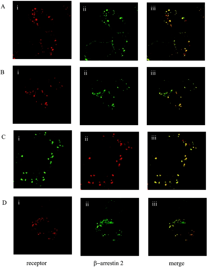
Figure 1. Orexin-1 receptor co-internalizes with β-arrestin-2.
HEK-293T cells were transfected with various forms of the human orexin-1 receptor and β-arrestin-2. Post-transfection (24 h) cells were challenged with orexin-A (0.5 μM) for 30 min. Cells were then fixed and visualized. The distribution of the orexin-1 receptor (i), β-arrestin-2 (ii) and a composite of these images (iii) are shown. (A) Wild-type orexin-1 receptor+β-arrestin-2–GFP, stimulated with TAMRA-orexin-A. (B) N-terminally HA-tagged orexin-1 receptor+β-arrestin-2–GFP, stimulated with orexin-A in the presence of anti-HA antibody. (C) Orexin-1 receptor–eYFP+β-arrestin-2–RFP, stimulated with orexin-A. (D) N-terminally VSV-G-tagged orexin-1 receptor+β-arrestin-2–GFP, stimulated with orexin-A in the presence of CypHer-5-labelled anti-VSV-G antibody.

