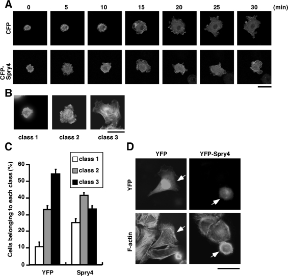
Figure 5. Spry4 suppresses cell spreading.
(A) Time-lapse analyses of the spreading of C2C12 cells after replating on laminin. Cells transfected with CFP or CFP–Spry4 together with YFP–actin were cultured for 18 h, suspended and replated on laminin-coated coverglasses. Cells were analysed by time-lapse fluorescence microscopy, making use of YFP fluorescence. Scale bar, 40 μm. (B) Three categories of spreading cells. C2C12 cells transfected with YFP or YFP–Spry4 were cultured and replated on laminin-coated coverglasses. After incubation for 30 min, cells were fixed and stained with rhodamine–phalloidin for F-actin. On the basis of the area of spreading cells, cells were categorized into three classes; cell area<800 μm2 (class 1), 800 μm2<cell area<1600 μm2 (class 2) and cell area>1600 μm2 (class 3). Scale bar, 40 μm. (C) Quantitative analysis of the effects of Spry4 expression on the spreading of C2C12 cells. Cells transfected with YFP or YFP–Spry4 were cultured for 18 h, suspended and replated on laminin-coated coverglasses. After incubation for 30 min, cells were fixed and stained with rhodamine–phalloidin. Cells were classified into three categories and percentages of these cells in YFP-positive cells (at least 200 cells) were calculated. The results are the means±S.E.M. for triplicate experiments. (D) Cell spreading morphologies of YFP- or YFP–Spry4-expressing cells. C2C12 cells transfected with YFP or YFP–Spry4 were replated on laminin and cultured for 30 min. Cells were fixed and analysed by YFP fluorescence (upper panels) and staining with rhodamine–phalloidin for F-actin (lower panels). Arrows indicate the YFP-positive cells. Scale bar, 40 μm.

