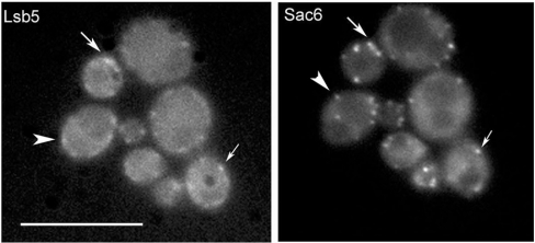
Figure 3. Co-localization of Lsb5p and Sac6p.
Yeast strain KAY446 expressing a GFP–Lsb5 plasmid was crossed with KAY684 carrying integrated Sac6–RFP, as described in the Materials and methods section. Arrows indicate regions of overlap of the staining patterns, indicating some co-localization. Other spots of Lsb5p or Sac6p fluorescence are clearly not overlapping (arrowheads), indicating that they exist in distinct complexes at the cell cortex. Scale bar, 10 μm.

