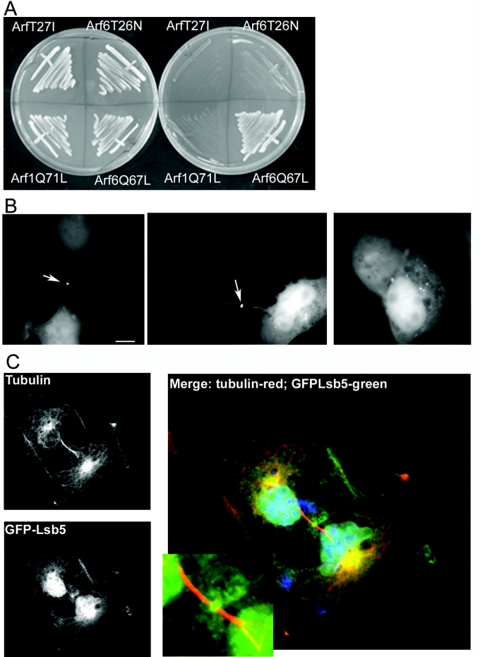
Figure 6. Interaction between Lsb5p and mammalian Arf6.
(A) Yeast two-hybrid analysis in cells expressing Lsb5 bait plasmid and different Arf constructs fused to an activation domain sequence. Only co-expression of Lsb5 and activated Arf6Q67L gave a positive interaction on selection plates. (B) GFP–Lsb5 was transfected into COS7 cells as described. Localization was observed in two main localizations: in punctate spots dispersed in the cytoplasm (right-hand panel) and at the midbody, indicated by arrows (left-hand and central panels). (C) To demonstrate that the staining of Lsb5GFP is at the midbody, we used immunofluorescence microscopy to observe tubulin localization in the cells. Tubulin localizes to distinct structures either side of a midbody, but has a clear zone at the midbody itself. As depicted, GFP–Lsb5p localizes to the midbody, as defined by the tubulin structures on either side. Scale bar, 10 μm.

