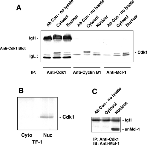
Figure 3. Co-immunoprecipitation results for Cdk1, Mcl-1 and snMcl-1.
(A) Cdk1 co-immunoprecipitates with Mcl-1. Cytosolic and nuclear proteins were extracted from asynchronous HL-60 cells and immunoprecipitated with antibody to Cdk-1, cyclin B1 or Mcl-1. Cdk1 (34 kDa) was detected by immunoblotting. IgH and IgL refer to bands detected due to immunoglobulin heavy and light chains respectively. (B) Co-immunoprecipitation of Mcl-1 and Cdk1 can be detected in TF-1 cells. Cytosolic (Cyto) and nuclear (Nuc) extracts from TF-1 cells were immunoprecipitated with anti-Mcl-1 and probed for the presence of Cdk1. The band below Cdk1 in (A) that was attributed to IgL was not seen in (B) due to the use of different batches of antibody. (C) snMcl-1 co-immunoprecipitates with anti-Cdk1. Cytosolic and nuclear proteins were extracted from asynchronous HL-60 cells and immunoprecipitated with anti-Cdk1. Immunoblotting with anti-Mcl-1 revealed a doublet at 35/36 kDa in the nuclear extract only that corresponds to snMcl-1. Each panel is representative of at least four independent experiments.

