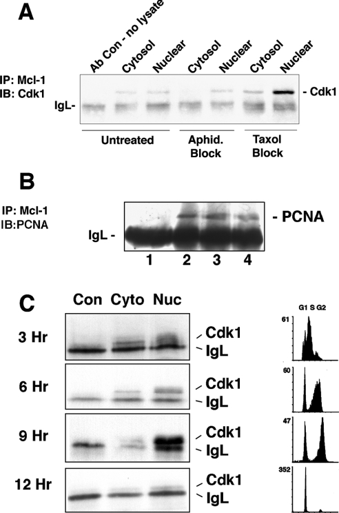
Figure 5. Co-immunoprecipitation of Mcl-1 and Cdk1 is maximal near G2/M.
(A) Cytosolic and nuclear proteins were extracted from HL-60 cells that were untreated, or treated with aphidicolin (Aphid.) to block cells at G1/S, or Taxol to block cells at G2/M. Presence of Cdk1 associated with Mcl-1 was determined. (B) HL-60 cells were either untreated (lane 2) or treated with aphidicolin (lane 3) or Taxol (lane 4). Lane 1 represents a mock immunoprecipitation with no cell lysate to show immunoglobulin light chain (IgL) and the other samples were immunoprecipitated with anti-Mcl-1. Immunoblotting was with antibody directed to PCNA. (C) HL-60 cells blocked with aphidicolin were washed and released for indicated times, at which point cytosolic (Cyto) and nuclear (Nuc) extracts were immunoprecipitated with anti-Mcl-1. Immunoblotting was done to detect Cdk1. Con indicates samples of a mock immunoprecipitation with the same antibody but no cell proteins. Flow cytometry was performed after PI staining to determine the cell-cycle stage at each of the time points. Results are representative of at least three independent experiments.

