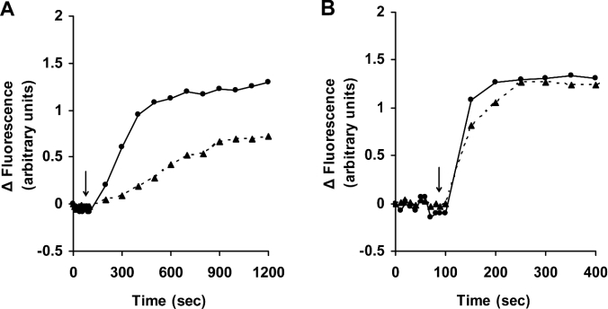
Figure 3. Iron chelation by DFO is temperature-dependent.
Jurkat cells (1.5×10⁶ cells/ml) were loaded with calcein as described for Figure 1. Cells were then collected, washed, resuspended in PBS containing 20 mM Hepes and 1 mg/ml BSA, and transferred to cuvettes maintained at either 37 °C (●) or 4 °C (▲), and fluorescence (excitation 488 nm; emission 517 nm) was monitored. At the time points indicated by the arrows, 1 mM DFO (A) or 11 μM SIH (B) was added directly into the cuvette, and the increase in fluorescence, indicating relocation of iron from calcein, was assayed. Two similar experiments gave essentially the same results.

