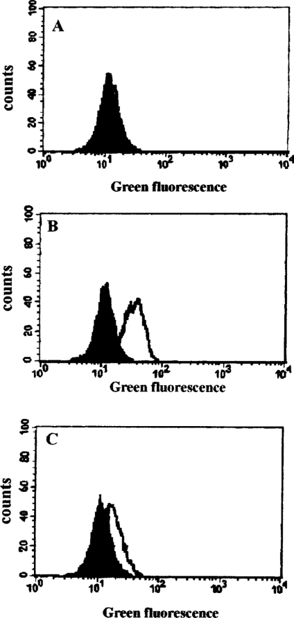
Figure 5. DFO inhibits H2O2-induced lysosomal membrane destabilization.
Jurkat cells (1.5×10⁶ cells/ml) were loaded with 0.1 μg/ml AO for 15 min before being collected and analysed for green fluorescence by flow cytometry (filled histograms in A, B and C). Samples from the same suspension of cells were also treated with 1 mM DFO for 2 h (open histogram, A), or oxidatively stressed by exposure to 0.1 μg/ml GO (generating approx. 2 nmol of H2O2/min per ml) for 1 h in complete medium (open histogram, B), or treated with 1 mM DFO for 2 h before exposure to the same amount of GO for a further 1 h (open histogram, C). The increase in green fluorescence in (B) indicates lysosomal destabilization with release of AO into the cytosol. The data shown are from one of three experiments giving essentially the same results.

