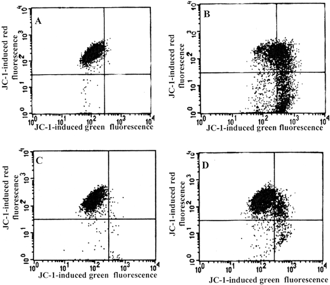
Figure 6. DFO protects against H2O2-induced loss of mitochondrial membrane potential.
Suspended Jurkat cells (1.5×10⁶ cells/ml) were exposed (B and D) or not (A and C) to continuously generated H2O2 for 5 h (0.1 μg/ml GO, able to generate approx. 2 nmol of H2O2/min per ml, was added to complete medium). In (C) and (D), cells were initially exposed to 1 mM DFO for 2 h before oxidative stress was generated. After exposure to oxidative stress, cells were collected by centrifugation, washed in PBS and resuspended for 15 min in PBS containing 1 μg/ml JC-1, and both green and red fluorescence were analysed by flow cytometry. Experiments were performed twice with essentially the same results.

