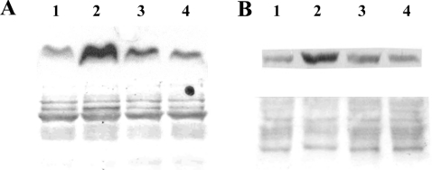
Figure 7. DFO protects against H2O2-induced release of cytochrome c and AIF from mitochondria.
Jurkat cells (1.5×10⁶ cells/ml), treated or not with 1 mM DFO for 2 h, were exposed to 0.1 μg/ml GO (generating approx. 2 nmol of H2O2/min per ml) for 5 h. Subsequently, cells were fractionated, and cytosolic (A) and nuclear (B) portions were isolated and analysed for the presence of cytochrome c and AIF respectively by Western blotting (see the Materials and methods section for details). The lower panels represent Ponceau staining of the relevant part of the membrane to show equal loading. Lane 1, untreated cells; lane 2, cells exposed to GO for 5 h; lane 3, cells pretreated with 1 mM DFO for 2 h before exposure to GO; lane 4, cells treated with DFO only for 7 h.

