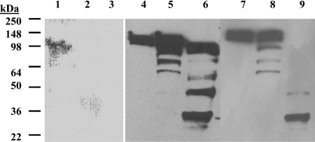
Figure 1. Expression of full-length mouse TACE wild-type and variants.
Western blot of mTACEΔPro (lanes 1, 2 and 3), wild-type (lanes 4, 5 and 6) and C184A (lanes 7, 8 and 9) forms. Cell-associated fractions were analysed at 24 h (lanes 1, 4 and 7), 48 h (lanes 2 and 5) and 72 h (lanes 3, 6 and 9) post-infection. Cell associated fractions were obtained after protein extraction with 1% NP40, followed by centrifugation, SDS/PAGE separation and Western blotting using ECL® for TACE polypeptide visualization (see the Materials and methods section). The mTACEΔPro signal was weaker and the gel was overdeveloped, resulting in higher noise levels.

