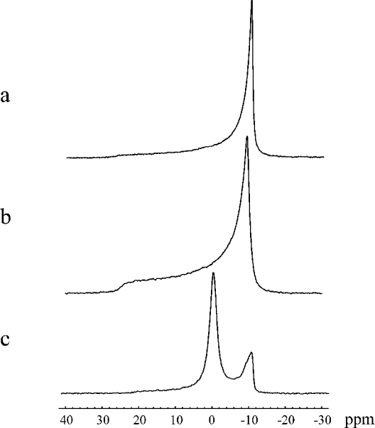
Figure 2. 31P-NMR spectra of MLV DPPG at 50 °C in the absence of CTI (a) and in the presence of CTI at L/P ratios of 40:1 (b) and 10:1 (c).
The parameters of CSA (Δσ) at the 31P nucleus of DPPG bilayer, the deformation (c/a) of the liposomes in the magnetic field and the content of isotropic signal estimated by computer simulation of the line shape were found to be respectively: (a) 38±1 p.p.m., 1.90±0.05, 0%; (b) 35±1 p.p.m., 1.49±0.05, 0% and (c) 30±1 p.p.m., 1.15±0.05, 78±1%.

