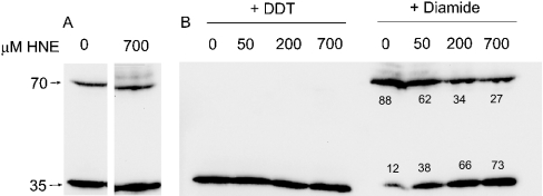
Figure 2. Immunodetection of Aox proteins from HNE-treated mitochondria of Arabidopsis cell cultures.
Isolated mitochondria (100 μl) were suspended in 1.25 of ml of reaction medium and treated with 0–700 μM HNE as indicated, for 20 min at 25 °C. Mitochondria were pelleted and re-suspended in 100 μl of wash buffer (without BSA). Samples (50 μg) were separated by SDS/PAGE, transferred on to a nitrocellulose membrane and probed with an anti-Aox antibody. (A) No further treatment; (B) treated with either 200 mM diamide or 20 mM DTT for 30 min at room temperature immediately prior to separation, as indicated. The positions of the 70- and 35-kDa molecular-mass markers are indicated on the left. Numbers under the bands represent the percentage of Aox as a dimer or monomer at each concentration of HNE.

