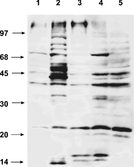
Figure 6. Immunodetection of mitochondrial proteins modified by HNE in oxidative stress-treated cells.
Proteins from mitochondria isolated from cell cultures, treated for 16 h with either 88 mM H2O2 (lane 3), 25 μM antimycin A (lane 4) or 400 μM menadione (lane 5), were separated by SDS/PAGE, transferred on to nylon membranes and probed with polyclonal anti-HNE-adduct antibodies. Control mitochondria from untreated cells (lane 1) and mitochondria treated directly with 700 μM HNE (lane 2) are also shown. The positions of molecular-mass markers are indicated on the left in kDa.

