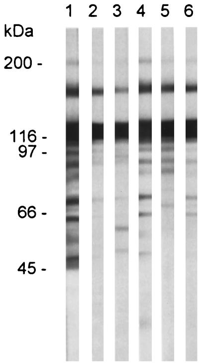
FIG. 4.
Reactivity of MSG-specific MAbs with P. carinii f. sp. carinii proteins. P. carinii f. sp. carinii proteins were separated by electrophoresis through an 8 to 16% Novex SDS-PAGE gel. Each lane contained lysate from approximately 107 organisms. Proteins were transferred to a nitrocellulose sheet, strips of which were incubated with the following mouse MAbs: lane 1, RA-E7; lane 2, RA-C1; lane 3, RA-C6; lane 4, RA-C7; lane 5, RB-C8; lane 6, RA-C11. Bound antibodies were detected as described in the legend to Fig. 3. Positions of molecular mass markers are indicated to the left of lane 1.

