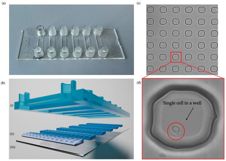
Figure 4.
Microfluidic chip with microwells. (a) Picture of a microfluidic chip with microwells with six Ibidi channels. (b) Schematic cross-section of (i) Ibidi chip with channels with inlet and outlet connections, (ii) PDMS membranes with microwells size 50 × 50 × 50 µm, and (iii) coverslip glass. (c) Brightfield image of PDMS membrane in the microfluidic chip. (d) Bright-field image of the microwell with a single C. albicans cell.

